Copyright
©2013 Baishideng Publishing Group Co.
World J Gastroenterol. Jun 7, 2013; 19(21): 3173-3188
Published online Jun 7, 2013. doi: 10.3748/wjg.v19.i21.3173
Published online Jun 7, 2013. doi: 10.3748/wjg.v19.i21.3173
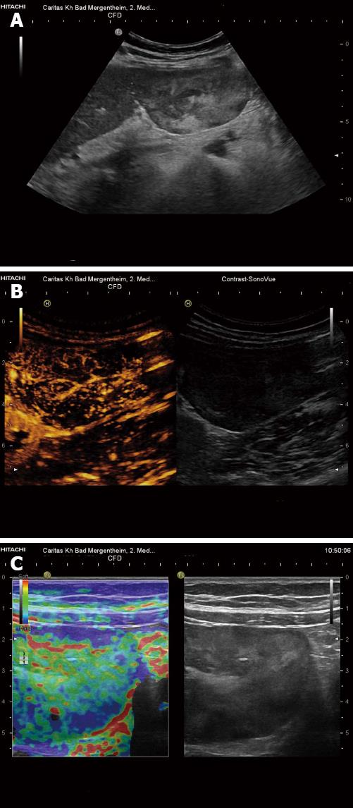
Figure 3 Teleangiectatic focal nodular hyperplasia.
Pedunculated liver tumor, histologically teleangiectatic focal nodular hyperplasia with signs of peliosis. A: B-mode imaging shows heterogenous echogenicity; B: Contrast-enhanced ultrasound reveals central arterial blood supply; C: Real-time elastography shows harder periphery and softer central portions of the lesion.
- Citation: Dietrich CF, Sharma M, Gibson RN, Schreiber-Dietrich D, Jenssen C. Fortuitously discovered liver lesions. World J Gastroenterol 2013; 19(21): 3173-3188
- URL: https://www.wjgnet.com/1007-9327/full/v19/i21/3173.htm
- DOI: https://dx.doi.org/10.3748/wjg.v19.i21.3173


