Copyright
©2013 Baishideng Publishing Group Co.
World J Gastroenterol. May 28, 2013; 19(20): 3027-3042
Published online May 28, 2013. doi: 10.3748/wjg.v19.i20.3027
Published online May 28, 2013. doi: 10.3748/wjg.v19.i20.3027
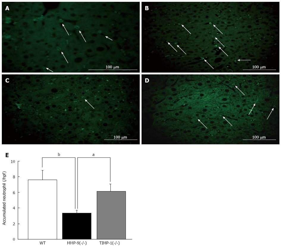
Figure 7 Immunohistochemistry analysis for accumulated neutrophils with myeloperoxidase staining between wild-type, matrix metalloproteinase-9(-/-) and tissue inhibitor of metalloprote-inase-1(-/-) mice 6 h after 80%-partial hepatectomy was performed.
Myeloperoxidase (MPO) staining labeled in green (Alexa Fluor 488) is shown (A). Cytoplasmic azurophilic granules were characteristically stained in neutrophils. Representative images of MPO staining on a remnant liver (RL) section in green in wild-type (WT) (B), matrix metalloproteinase-9 (MMP-9)(-/-) (C), tissue inhibitor of metalloproteinase-1 (TIMP-1)(-/-) mice (D) are shown. A histogram of the number of accumulated neutrophils in the liver is shown. We observed significantly fewer neutrophils in the RL in MMP-9(-/-) mice than in WT and TIMP-1(-/-) mice (aP < 0.05, bP < 0.01 vs WT and TIMP-1) (E).
- Citation: Ohashi N, Hori T, Chen F, Jermanus S, Nakao A, Uemoto S, Nguyen JH. Matrix metalloproteinase-9 in the initial injury after hepatectomy in mice. World J Gastroenterol 2013; 19(20): 3027-3042
- URL: https://www.wjgnet.com/1007-9327/full/v19/i20/3027.htm
- DOI: https://dx.doi.org/10.3748/wjg.v19.i20.3027


