Copyright
©2013 Baishideng Publishing Group Co.
World J Gastroenterol. May 28, 2013; 19(20): 3027-3042
Published online May 28, 2013. doi: 10.3748/wjg.v19.i20.3027
Published online May 28, 2013. doi: 10.3748/wjg.v19.i20.3027
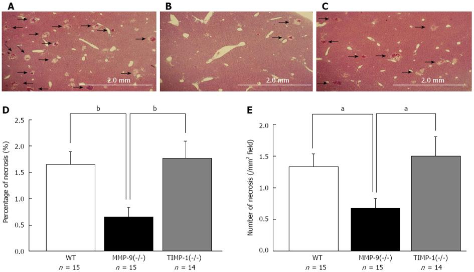
Figure 5 Histological analysis between wild-type, matrix metalloproteinase-9(-/-), and tissue inhibitor of metalloproteinase-1(-/-) mice 6 h after 80%-partial hepatectomy were performed.
Representative images of the remnant liver (RL) 6 h after 80%-PH in wild-type (WT) (A), matrix metalloproteinase-9 (MMP-9)(-/-) (B), and tissue inhibitor of metalloproteinase-1 (TIMP-1)(-/-) mice (C) are shown. Samples that included necrosis were selected for this figure. The percentage (D) and the number of necrotic foci per mm2 (E) of the necrotic area in the RL are shown. We observed significantly smaller and less necrotic foci in MMP-9(-/-) mice compared with WT and TIMP-1(-/-) mice (aP < 0.05, bP < 0.01 vs WT and TIMP-1).
- Citation: Ohashi N, Hori T, Chen F, Jermanus S, Nakao A, Uemoto S, Nguyen JH. Matrix metalloproteinase-9 in the initial injury after hepatectomy in mice. World J Gastroenterol 2013; 19(20): 3027-3042
- URL: https://www.wjgnet.com/1007-9327/full/v19/i20/3027.htm
- DOI: https://dx.doi.org/10.3748/wjg.v19.i20.3027


