Copyright
©2013 Baishideng Publishing Group Co.
World J Gastroenterol. May 28, 2013; 19(20): 3027-3042
Published online May 28, 2013. doi: 10.3748/wjg.v19.i20.3027
Published online May 28, 2013. doi: 10.3748/wjg.v19.i20.3027
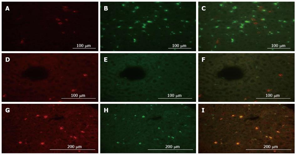
Figure 4 Localization analysis of matrix metalloproteinase-9 by dual immune-fluorescence in the remnant liver after 80%-partial hepatectomy is shown.
A: Fluorescent microscopy images of matrix metalloproteinase-9 (MMP-9) labeled in red (Alexa Fluor 568); B: CD68 labeled in green (Alexa Fluor 488); C: A merged image of MMP-9 and CD68 staining are shown; D: MMP-9 labeled in red (Alexa Fluor 568); E: Desmin labeled in green (Alexa Fluor 488); and F: A merged image of MMP-9 and desmin are shown; G: MMP-9 labeled in red (Alexa Fluor 568); H: CD11b labeled in green (Alexa Fluor 488); I: A merged image of MMP-9 and CD11b are shown. Protein expression of MMP-9 was mainly localized in CD11b-positive cells and desmin-positive cells to some degree.
- Citation: Ohashi N, Hori T, Chen F, Jermanus S, Nakao A, Uemoto S, Nguyen JH. Matrix metalloproteinase-9 in the initial injury after hepatectomy in mice. World J Gastroenterol 2013; 19(20): 3027-3042
- URL: https://www.wjgnet.com/1007-9327/full/v19/i20/3027.htm
- DOI: https://dx.doi.org/10.3748/wjg.v19.i20.3027


