Copyright
©2013 Baishideng Publishing Group Co.
World J Gastroenterol. May 28, 2013; 19(20): 3027-3042
Published online May 28, 2013. doi: 10.3748/wjg.v19.i20.3027
Published online May 28, 2013. doi: 10.3748/wjg.v19.i20.3027
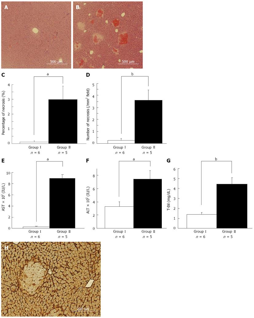
Figure 1 Necrosis is a pivotal event for postoperative liver failure in the remnant liver after 80%-partial hepatectomy.
There were histological and hematological differences between asymptomatic and symptomatic mice suspected as having postoperative liver failure 6 h after 80%-partial hepatectomy (PH) (A, B). Representative images of the remnant liver (RL) 6 h after 80%-PH in asymptomatic mice (A) and symptomatic mice (B) are shown. Significant multiple necroses with microhemorrhage were observed in the middle zone in symptomatic mice (B), while no obvious liver damage was observed in asymptomatic mice (A). The percentage area of necrosis (C) and the number of necrosis in each mm2 (D) in the RL were confirmed. Necrotic areas were significantly larger and more prevalent in symptomatic mice liver compared with asymptomatic mice. Serum levels of aspartate aminotransferase (AST) (E), alamine aminotransferase (ALT) (F) and total bilirubin (T-Bil) (G) 6 h after 80%-PH are shown. AST, ALT and T-Bil levels were significantly elevated in symptomatic mice compared with those in asymptomatic mice. Representative image of immunohistochemistry of intercellular adhesion molecule-1 (ICAM-1) in the RL 6 h after 80%-PH is shown (H). ICAM-1 expression was clearly observed, particularly in the sinusoid lining in areas without necrosis. We observed a breakdown in sinusoid structure with hemorrhage in necrotic areas after 80%-PH in mice, aP < 0.05, bP < 0.01 vs asymptomatic mice.
- Citation: Ohashi N, Hori T, Chen F, Jermanus S, Nakao A, Uemoto S, Nguyen JH. Matrix metalloproteinase-9 in the initial injury after hepatectomy in mice. World J Gastroenterol 2013; 19(20): 3027-3042
- URL: https://www.wjgnet.com/1007-9327/full/v19/i20/3027.htm
- DOI: https://dx.doi.org/10.3748/wjg.v19.i20.3027


