Copyright
©2013 Baishideng Publishing Group Co.
World J Gastroenterol. Jan 14, 2013; 19(2): 299-303
Published online Jan 14, 2013. doi: 10.3748/wjg.v19.i2.299
Published online Jan 14, 2013. doi: 10.3748/wjg.v19.i2.299
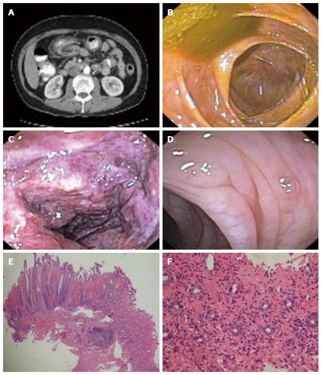
Figure 1 The laboratory studies results.
A: Computed tomography scan shows thickening of the colonic wall involving the splenic flexure of the colon; B: Normal mucosa in the right colon (appendiceal orifice); C: Ischemic colitis features in the splenic flexure of the colon; D: Normal mucosa in sigmoid with a small polyp; E, F: Histopathology shows ischemic loss of crypts and acute hemorrhage in lamnia propria (acute hemorrhagic necrosis replacing glands) associated with acute fibrinopurulent exudate at colonic epithelial surface, consistent with ischemic colitis.
- Citation: Sherid M, Sifuentes H, Samo S, Deepak P, Sridhar S. Lubiprostone induced ischemic colitis. World J Gastroenterol 2013; 19(2): 299-303
- URL: https://www.wjgnet.com/1007-9327/full/v19/i2/299.htm
- DOI: https://dx.doi.org/10.3748/wjg.v19.i2.299


