Copyright
©2013 Baishideng Publishing Group Co.
World J Gastroenterol. Jan 14, 2013; 19(2): 161-164
Published online Jan 14, 2013. doi: 10.3748/wjg.v19.i2.161
Published online Jan 14, 2013. doi: 10.3748/wjg.v19.i2.161
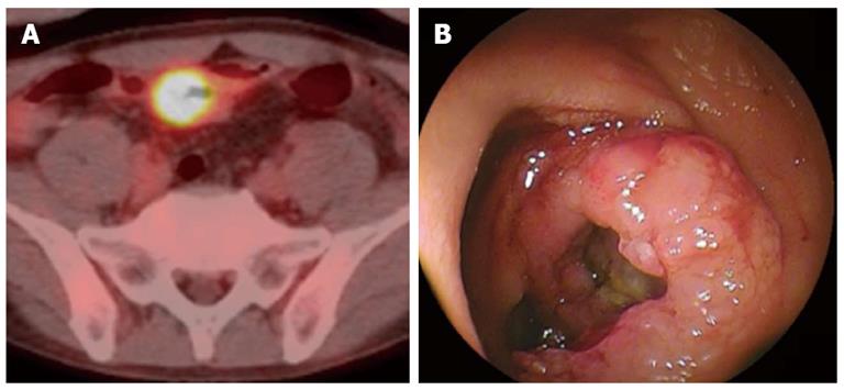
Figure 2 A mass positive by positron emission computed tomography actually was a tumor arising the mucosa of the small intestine of a 48-year-old woman who had suffered Crohn disease.
A: Positron emission computed tomography/computed tomography showed an accumulation in site of wall thickening of ileum; B: Image obtained by double-balloon enteroscopy.
- Citation: Sugimura H, Osawa S. Internal frontier: The pathophysiology of the small intestine. World J Gastroenterol 2013; 19(2): 161-164
- URL: https://www.wjgnet.com/1007-9327/full/v19/i2/161.htm
- DOI: https://dx.doi.org/10.3748/wjg.v19.i2.161


