Copyright
©2013 Baishideng Publishing Group Co.
World J Gastroenterol. May 7, 2013; 19(17): 2714-2717
Published online May 7, 2013. doi: 10.3748/wjg.v19.i17.2714
Published online May 7, 2013. doi: 10.3748/wjg.v19.i17.2714
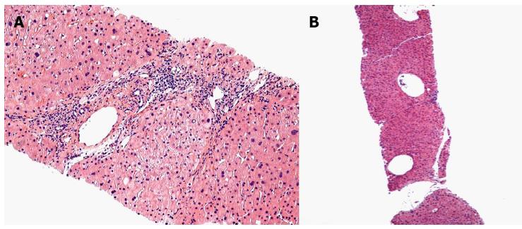
Figure 2 The hepatic histology of arterioportal fistula.
A: This low power image shows a small portal tract in the center of the field that has a markedly dilated portal venule. The terminal hepatic (central) venules above and below the portal tract are markedly dilated; B: The portal venule is slightly dilated and there are increased numbers of small portal venous radicals.
- Citation: Nookala A, Saberi B, Ter-Oganesyan R, Kanel G, Duong P, Saito T. Isolated arterioportal fistula presenting with variceal hemorrhage. World J Gastroenterol 2013; 19(17): 2714-2717
- URL: https://www.wjgnet.com/1007-9327/full/v19/i17/2714.htm
- DOI: https://dx.doi.org/10.3748/wjg.v19.i17.2714


