Copyright
©2013 Baishideng Publishing Group Co.
World J Gastroenterol. May 7, 2013; 19(17): 2638-2649
Published online May 7, 2013. doi: 10.3748/wjg.v19.i17.2638
Published online May 7, 2013. doi: 10.3748/wjg.v19.i17.2638
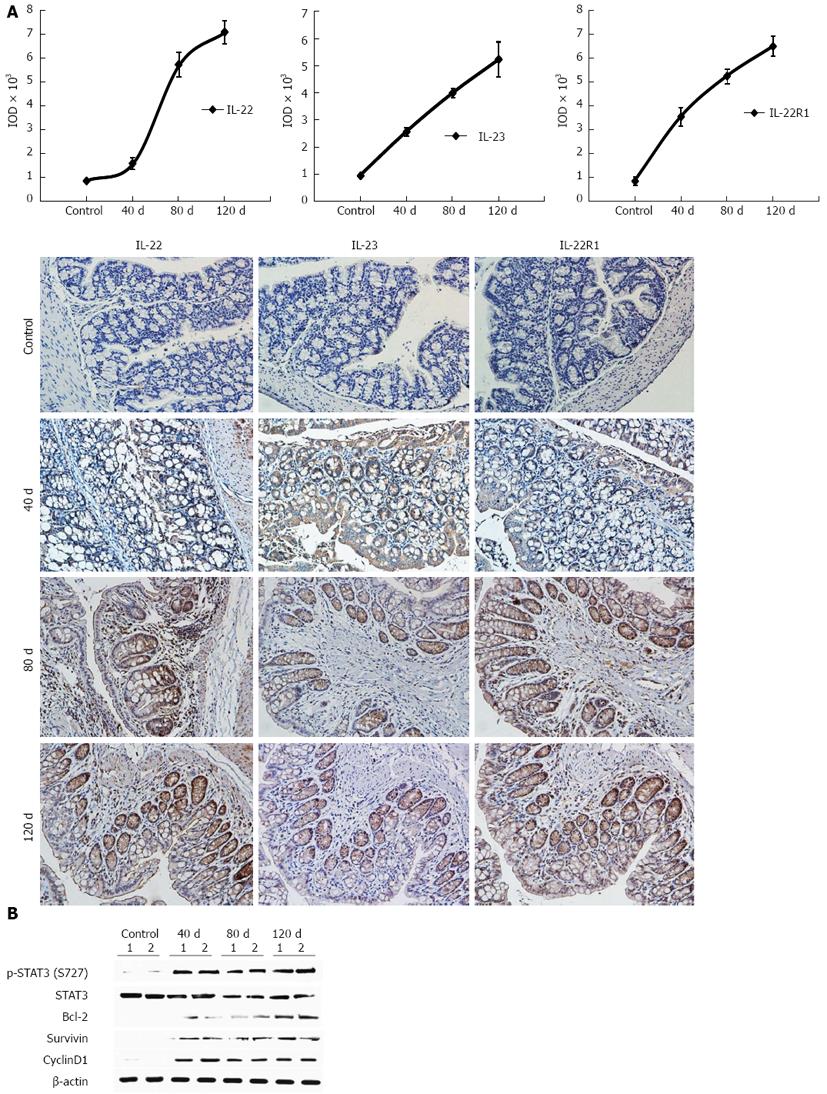
Figure 7 Expression of interleukin-22 and its related proteins in the mouse chronic colitis model.
A: The average integrated optical density (IOD) for the immunohistochemistry staining of interleukin (IL)-22, IL-23, and IL-22R1 in mouse ulcerative colitis and normal tissues at different time points: days 40, 80, and 120. The expression of IL-22, IL-22R1, and IL-23 gradually increased with time; B: Western blotting detection of p-STAT3 (S727), total STAT3, Bcl-2, survivin, and cyclin D1 after DSS administration. Expression levels were all normalized to β-actin. All genes showed sustained expression over time.
- Citation: Yu LZ, Wang HY, Yang SP, Yuan ZP, Xu FY, Sun C, Shi RH. Expression of interleukin-22/STAT3 signaling pathway in ulcerative colitis and related carcinogenesis. World J Gastroenterol 2013; 19(17): 2638-2649
- URL: https://www.wjgnet.com/1007-9327/full/v19/i17/2638.htm
- DOI: https://dx.doi.org/10.3748/wjg.v19.i17.2638


