Copyright
©2013 Baishideng Publishing Group Co.
World J Gastroenterol. May 7, 2013; 19(17): 2629-2637
Published online May 7, 2013. doi: 10.3748/wjg.v19.i17.2629
Published online May 7, 2013. doi: 10.3748/wjg.v19.i17.2629
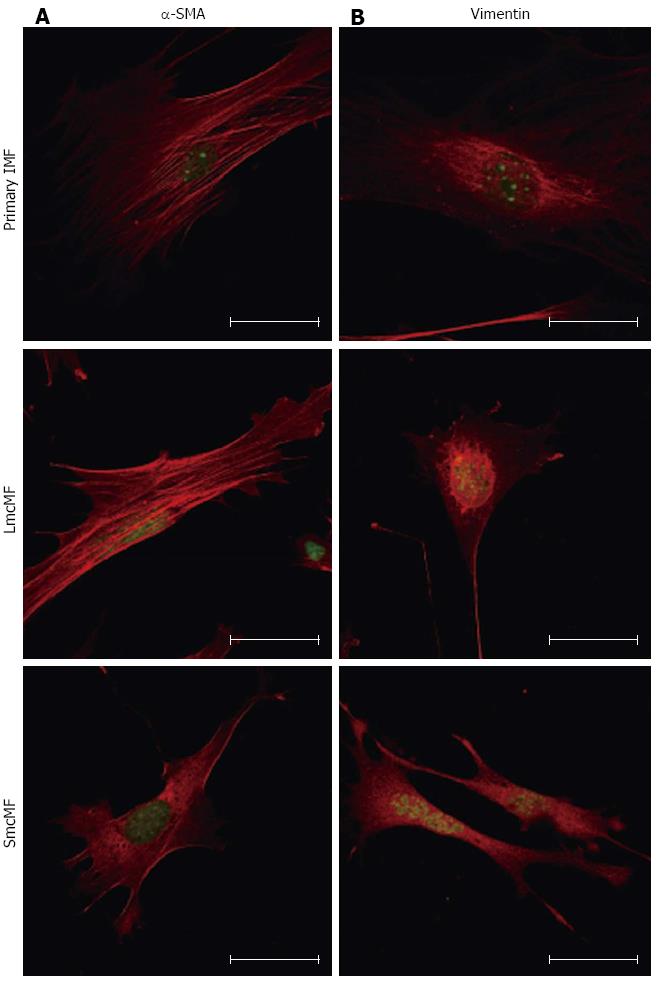
Figure 4 Immunofluorescence staining for α-smooth muscle actin and vimentin in primary intestinal myofibroblasts, LmcMF, and SmcMF.
Primary intestinal myofibroblasts (IMFs), LmcMF, and SmcMF were immunostained with α-smooth muscle actin (α-SMA) (A) and vimentin (B) (red). SYTOX Green was used for nuclear labeling (green). Representative images are shown. Scale bars indicate 40 μm.
- Citation: Kawasaki H, Ohama T, Hori M, Sato K. Establishment of mouse intestinal myofibroblast cell lines. World J Gastroenterol 2013; 19(17): 2629-2637
- URL: https://www.wjgnet.com/1007-9327/full/v19/i17/2629.htm
- DOI: https://dx.doi.org/10.3748/wjg.v19.i17.2629


