Copyright
©2013 Baishideng Publishing Group Co.
World J Gastroenterol. Apr 28, 2013; 19(16): 2578-2582
Published online Apr 28, 2013. doi: 10.3748/wjg.v19.i16.2578
Published online Apr 28, 2013. doi: 10.3748/wjg.v19.i16.2578
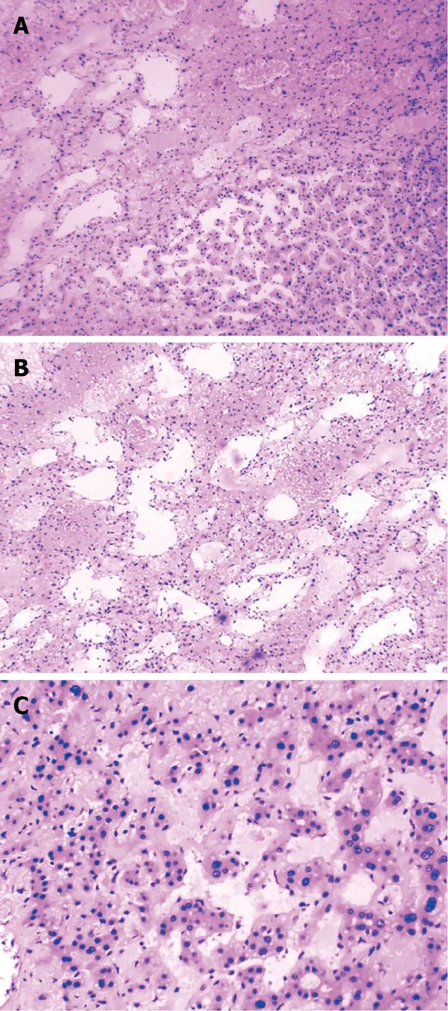
Figure 4 Pathologic examination, showing irregular and dilated lacunae, some of which were filled with blood and without endothelial linings.
Hematoxylin and eosin staining, original magnification A: × 100; B: × 200; C: × 400.
- Citation: Pan W, Hong HJ, Chen YL, Han SH, Zheng CY. Surgical treatment of a patient with peliosis hepatis: A case report. World J Gastroenterol 2013; 19(16): 2578-2582
- URL: https://www.wjgnet.com/1007-9327/full/v19/i16/2578.htm
- DOI: https://dx.doi.org/10.3748/wjg.v19.i16.2578


