Copyright
©2013 Baishideng Publishing Group Co.
World J Gastroenterol. Apr 28, 2013; 19(16): 2569-2573
Published online Apr 28, 2013. doi: 10.3748/wjg.v19.i16.2569
Published online Apr 28, 2013. doi: 10.3748/wjg.v19.i16.2569
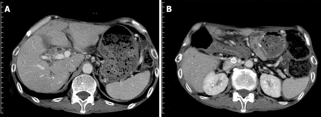
Figure 2 Computed tomography scan 3 mo after the expandable metallic stent placement.
A: Shows that the ascites decreased and the liver atrophy improved; B: The stent placed in the portal vein remained patent.
- Citation: Tsuruga Y, Kamachi H, Wakayama K, Kakisaka T, Yokoo H, Kamiyama T, Taketomi A. Portal vein stenosis after pancreatectomy following neoadjuvant chemoradiation therapy for pancreatic cancer. World J Gastroenterol 2013; 19(16): 2569-2573
- URL: https://www.wjgnet.com/1007-9327/full/v19/i16/2569.htm
- DOI: https://dx.doi.org/10.3748/wjg.v19.i16.2569


