Copyright
©2013 Baishideng Publishing Group Co.
World J Gastroenterol. Apr 21, 2013; 19(15): 2319-2330
Published online Apr 21, 2013. doi: 10.3748/wjg.v19.i15.2319
Published online Apr 21, 2013. doi: 10.3748/wjg.v19.i15.2319
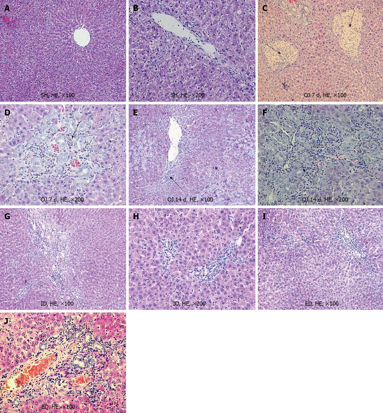
Figure 2 Micrograph of liver sections from rats of sham operation, obstructive jaundice, internal and external biliary drainage groups (the pathological changes were marked by black arrows).
A, B: There was almost no pathological changes in the liver of sham operation (SH) rats, the liver lobular architecture was intact; C, D: After bile duct ligation for 7 d, the liver showed focal necrosis and mild bile duct proliferation with acute inflammatory cell infiltration in the portal and periportal areas, but the lobular architecture was still intact in the cholestatic liver; E, F: After bile duct ligation for 14 d, the liver showed striking liver fibrosis and prominent bile duct proliferation in the portal tracts; G, H: Following 7 d of internal biliary drainage (ID), the liver displayed preserved lobular architecture with only mild bile duct proliferation, and the progression of liver fibrosis and cirrhosis stopped; I, J: After external biliary drainage (ED), lobular architecture was preserved with only mild biliary proliferation, but the degree of liver fibrosis was still at a high level. HE: Hematoxylin and eosin; OJ: Obstructive jaundice.
- Citation: Wang ZK, Xiao JG, Huang XF, Gong YC, Li W. Effect of biliary drainage on inducible nitric oxide synthase, CD14 and TGR5 expression in obstructive jaundice rats. World J Gastroenterol 2013; 19(15): 2319-2330
- URL: https://www.wjgnet.com/1007-9327/full/v19/i15/2319.htm
- DOI: https://dx.doi.org/10.3748/wjg.v19.i15.2319


