Copyright
©2013 Baishideng Publishing Group Co.
World J Gastroenterol. Mar 28, 2013; 19(12): 2005-2008
Published online Mar 28, 2013. doi: 10.3748/wjg.v19.i12.2005
Published online Mar 28, 2013. doi: 10.3748/wjg.v19.i12.2005
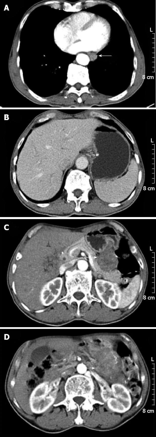
Figure 2 Computed tomography scan.
A: Circumferential thickening (arrow) of the lower esophageal wall with loss of lumen; B: Lower level displayed focal thickening (arrow) of the gastric wall with marked enhancement; C and D: Lower levels displayed a large, heterogeneous, round mass close to the greater curvature of the stomach (arrows).
- Citation: Zhou Y, Wu XD, Shi Q, Jia J. Coexistence of gastrointestinal stromal tumor, esophageal and gastric cardia carcinomas. World J Gastroenterol 2013; 19(12): 2005-2008
- URL: https://www.wjgnet.com/1007-9327/full/v19/i12/2005.htm
- DOI: https://dx.doi.org/10.3748/wjg.v19.i12.2005


