Copyright
©2013 Baishideng Publishing Group Co.
World J Gastroenterol. Mar 28, 2013; 19(12): 2000-2004
Published online Mar 28, 2013. doi: 10.3748/wjg.v19.i12.2000
Published online Mar 28, 2013. doi: 10.3748/wjg.v19.i12.2000
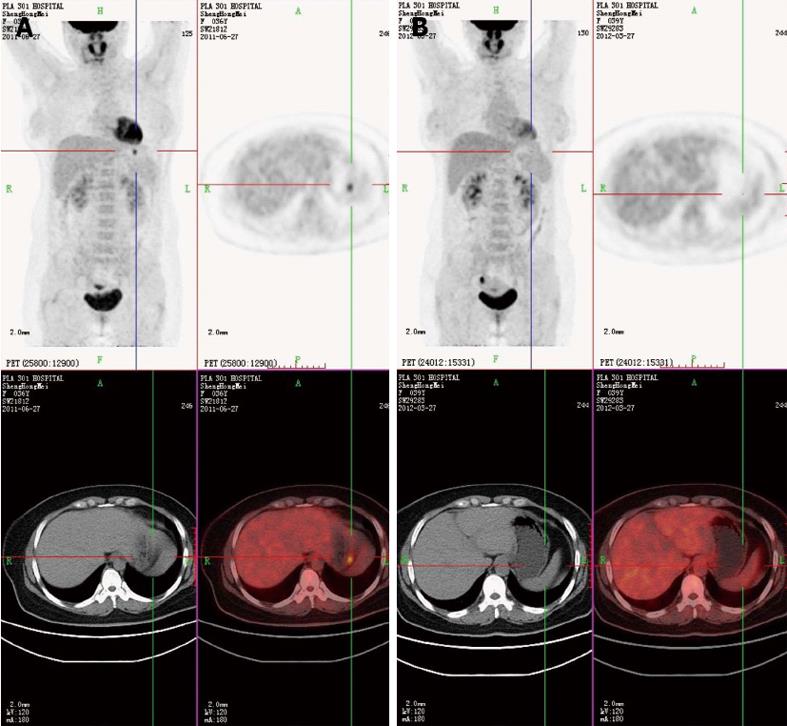
Figure 1 Positron emission tomography-computed tomography.
A: Positron emission tomography-computed tomography showed a nodular strong accumulation point with standardized uptake value 5.6 in the gastric fundus; B: After treatment, the nodular strong accumulation point in the gastric fundus had disappeared.
-
Citation: Li TT, Qiu F, Wang ZQ, Sun L, Wan J. Rare case of
Helicobacter pylori -related gastric ulcer: Malignancy or pseudomorphism? World J Gastroenterol 2013; 19(12): 2000-2004 - URL: https://www.wjgnet.com/1007-9327/full/v19/i12/2000.htm
- DOI: https://dx.doi.org/10.3748/wjg.v19.i12.2000


