Copyright
©2013 Baishideng Publishing Group Co.
World J Gastroenterol. Mar 28, 2013; 19(12): 1992-1996
Published online Mar 28, 2013. doi: 10.3748/wjg.v19.i12.1992
Published online Mar 28, 2013. doi: 10.3748/wjg.v19.i12.1992
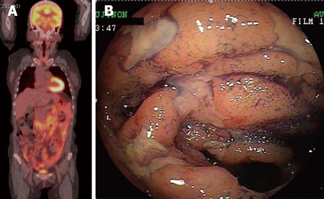
Figure 3 A 61-year-old woman with follicular lymphoma of the gastrointestinal tract.
A: 18F-fluorodeoxyglucose positron emission tomography combined with computed tomography in a projected image showed hypermetabolic foci in broad areas of the gastrointestinal tract from the 2nd portion of the duodenum to the terminal ileum (maximum standardized uptake value, 5.6); B: The colonoscopic view with indigo carmine dye-spray of the terminal ileum. Multiple ulcers with irregular margins were observed.
- Citation: Tari A, Asaoku H, Kunihiro M, Tanaka S, Yoshino T. Usefulness of positron emission tomography in primary intestinal follicular lymphoma. World J Gastroenterol 2013; 19(12): 1992-1996
- URL: https://www.wjgnet.com/1007-9327/full/v19/i12/1992.htm
- DOI: https://dx.doi.org/10.3748/wjg.v19.i12.1992


