Copyright
©2013 Baishideng Publishing Group Co.
World J Gastroenterol. Mar 28, 2013; 19(12): 1984-1991
Published online Mar 28, 2013. doi: 10.3748/wjg.v19.i12.1984
Published online Mar 28, 2013. doi: 10.3748/wjg.v19.i12.1984
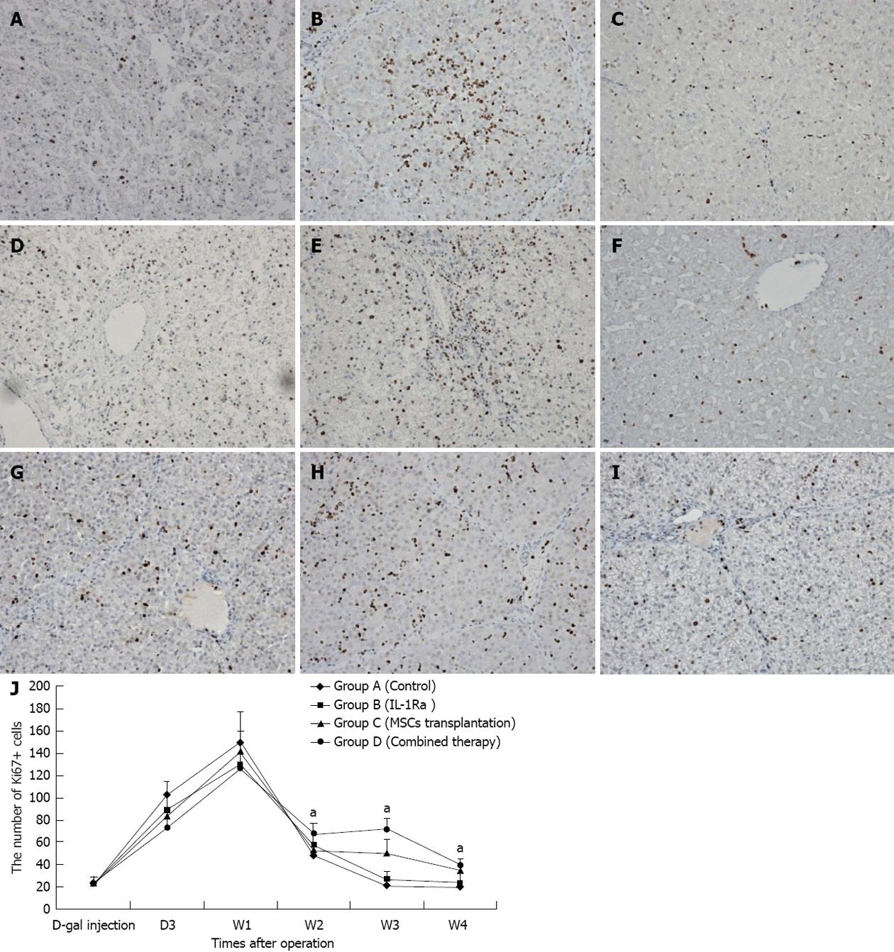
Figure 4 Changes in liver cell proliferation in all four groups.
A, B and C showed the anti-Ki67 stain of Group A at the time point of D3, W1 and W4, respectively (×200); D, E, F and G, H, I showed anti-Ki67 stain of Group B and Group C, respectively, just as Group A did (×200); J: Changes in the number of Ki67+ cells demonstrated that Group D had better long-term proliferation. aP < 0.05 vs control group. IL-1Ra: Interleukin-1 receptor antagonist; MSCs: Mesenchymal stem cells; D-gal: D-galactosamine.
- Citation: Shi XL, Zhu W, Tan JJ, Xiao JQ, Zhang L, Xu Q, Ma ZL, Ding YT. Effect evaluation of interleukin-1 receptor antagonist nanoparticles for mesenchymal stem cell transplantation. World J Gastroenterol 2013; 19(12): 1984-1991
- URL: https://www.wjgnet.com/1007-9327/full/v19/i12/1984.htm
- DOI: https://dx.doi.org/10.3748/wjg.v19.i12.1984


