Copyright
©2013 Baishideng Publishing Group Co.
World J Gastroenterol. Mar 21, 2013; 19(11): 1834-1840
Published online Mar 21, 2013. doi: 10.3748/wjg.v19.i11.1834
Published online Mar 21, 2013. doi: 10.3748/wjg.v19.i11.1834
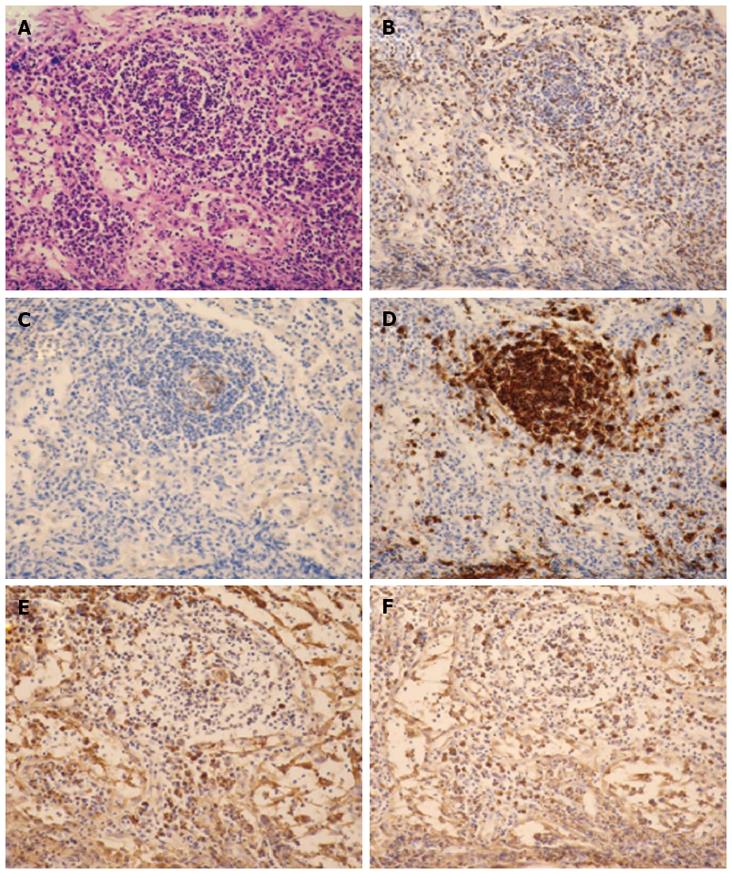
Figure 3 Histological sections of hepatoduodenal ligament lymph nodes (× 200 magnification).
A: The lymphoid follicles contain a reactive germinal center with hematoxylin and eosin staining. No evidences of granuloma or necrosis are visible; B: CD3 (T-cell marker) is positive in the interfollicular areas; C: CD10 (a marker of B-cell activation) is positive in the center of the follicles; D: CD20 (B-cell marker) is observed in the nodules; E: Kappa chain; F: Lambda chain. Neither the kappa nor the lambda chains predominate.
- Citation: Fujii H, Ohnishi N, Shimura K, Sakamoto M, Ohkawara T, Sawa Y, Nishida K, Ohkawara Y, Kobata T, Yamaguchi K, Itoh Y. Case of autoimmune hepatitis with markedly enlarged hepatoduodenal ligament lymph nodes. World J Gastroenterol 2013; 19(11): 1834-1840
- URL: https://www.wjgnet.com/1007-9327/full/v19/i11/1834.htm
- DOI: https://dx.doi.org/10.3748/wjg.v19.i11.1834


