Copyright
©2013 Baishideng Publishing Group Co.
World J Gastroenterol. Mar 21, 2013; 19(11): 1834-1840
Published online Mar 21, 2013. doi: 10.3748/wjg.v19.i11.1834
Published online Mar 21, 2013. doi: 10.3748/wjg.v19.i11.1834
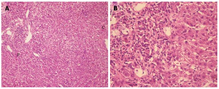
Figure 2 Liver biopsy.
A: Liver biopsy specimen with hematoxylin and eosin staining (× 100 magnification) revealing the histopathological appearance of acute hepatitis. Interface hepatitis and plasmacytic infiltrates are present; B: This is the same image at × 400 magnification. P: Portal area; C: Central vein area.
- Citation: Fujii H, Ohnishi N, Shimura K, Sakamoto M, Ohkawara T, Sawa Y, Nishida K, Ohkawara Y, Kobata T, Yamaguchi K, Itoh Y. Case of autoimmune hepatitis with markedly enlarged hepatoduodenal ligament lymph nodes. World J Gastroenterol 2013; 19(11): 1834-1840
- URL: https://www.wjgnet.com/1007-9327/full/v19/i11/1834.htm
- DOI: https://dx.doi.org/10.3748/wjg.v19.i11.1834


