Copyright
©2013 Baishideng Publishing Group Co.
World J Gastroenterol. Mar 21, 2013; 19(11): 1760-1769
Published online Mar 21, 2013. doi: 10.3748/wjg.v19.i11.1760
Published online Mar 21, 2013. doi: 10.3748/wjg.v19.i11.1760
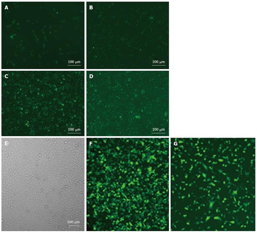
Figure 2 Transfection efficiency and cell morphology were detected by fluorescence microscope after adenovirus Class I phosphoinositide 3-kinase-RNA interference-green fluorescent protein and adenovirus negative control-RNA interference-green fluorescent protein treatment.
A-D: SGC7901 cells; E-G:MGC803 cells incubated with adenovirus Class I phosphoinositide 3-kinase [PI3K(I)]-RNA interference-green fluorescent protein (RNAi-GFP) (50 MOI) for the indicated time. A and E: Control group; B and F: 24 h after adenovirus PI3K(I)-RNAi-GFP treatment; C and G: 48 h adenovirus PI3K(I)-RNAi-GFP (50 MOI) treatment; D: 72 h after adenovirus PI3K(I)-RNAi-GFP (50 MOI) treatment (× 200) (n = 3).
- Citation: Zhu BS, Yu LY, Zhao K, Wu YY, Cheng XL, Wu Y, Zhong FY, Gong W, Chen Q, Xing CG. Effects of small interfering RNA inhibit Class I phosphoinositide 3-kinase on human gastric cancer cells. World J Gastroenterol 2013; 19(11): 1760-1769
- URL: https://www.wjgnet.com/1007-9327/full/v19/i11/1760.htm
- DOI: https://dx.doi.org/10.3748/wjg.v19.i11.1760


