Copyright
©2013 Baishideng Publishing Group Co.
World J Gastroenterol. Mar 14, 2013; 19(10): 1657-1660
Published online Mar 14, 2013. doi: 10.3748/wjg.v19.i10.1657
Published online Mar 14, 2013. doi: 10.3748/wjg.v19.i10.1657
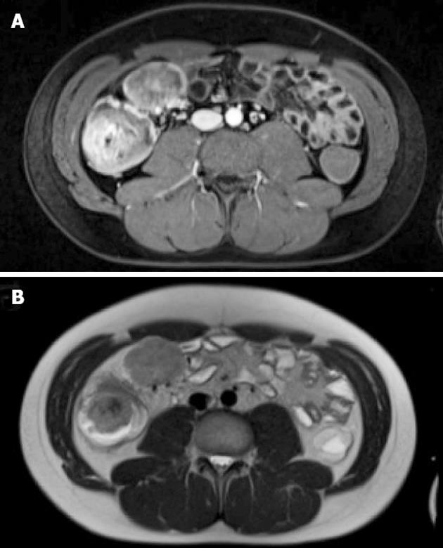
Figure 2 Magnetic resonance imaging of cecal perivascular epithelioid cell tumor and mesenteric lymph node metastasis.
A: T2-weighted image; B: T1-weighted image after iv administration of contrast medium.
- Citation: Scheppach W, Reissmann N, Sprinz T, Schippers E, Schoettker B, Mueller JG. PEComa of the colon resistant to sirolimus but responsive to doxorubicin/ifosfamide. World J Gastroenterol 2013; 19(10): 1657-1660
- URL: https://www.wjgnet.com/1007-9327/full/v19/i10/1657.htm
- DOI: https://dx.doi.org/10.3748/wjg.v19.i10.1657


