Copyright
©2013 Baishideng Publishing Group Co.
World J Gastroenterol. Mar 14, 2013; 19(10): 1582-1592
Published online Mar 14, 2013. doi: 10.3748/wjg.v19.i10.1582
Published online Mar 14, 2013. doi: 10.3748/wjg.v19.i10.1582
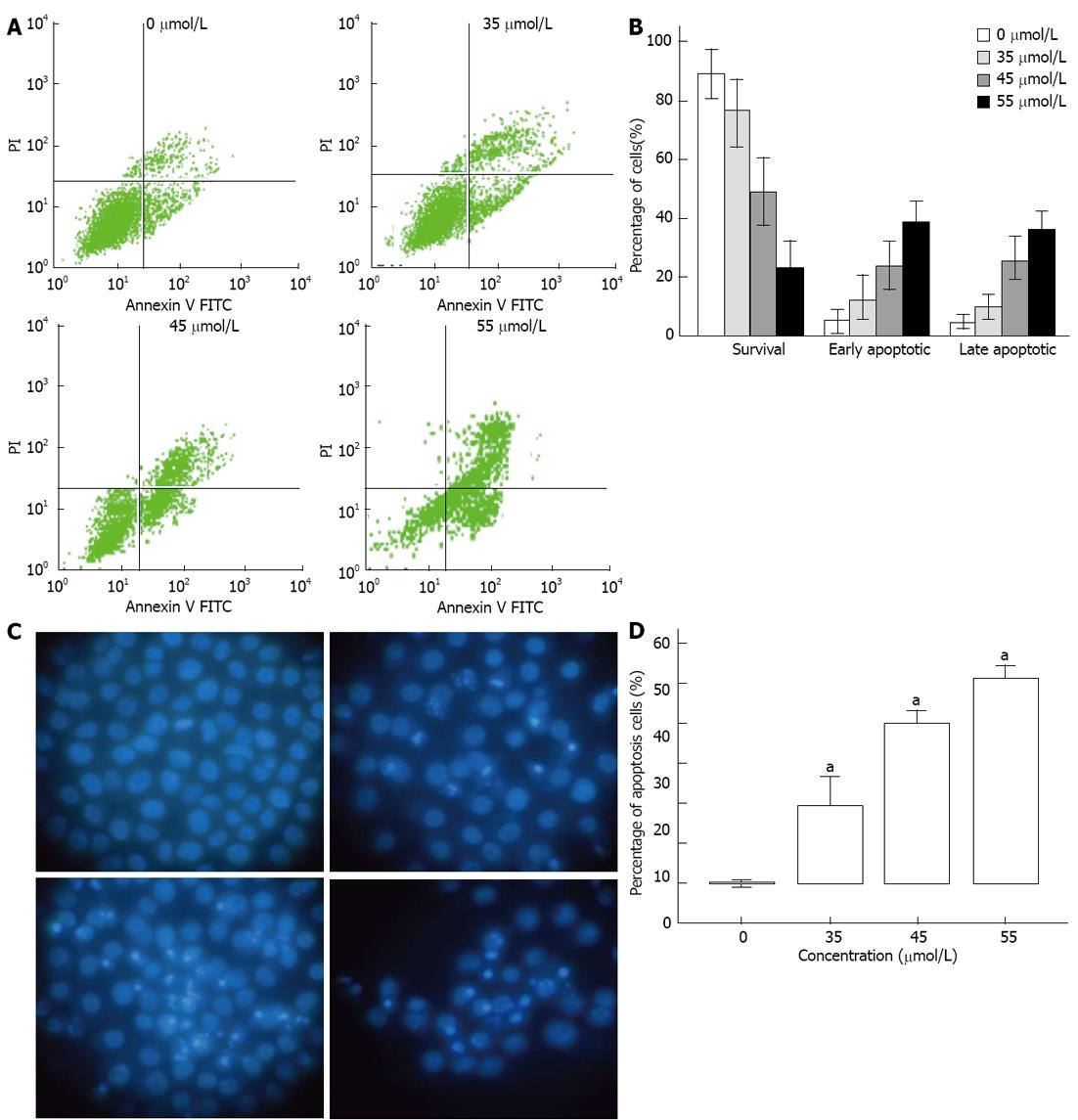
Figure 3 Rh2 induces Bxpc-3 cells apoptosis.
A, B: An apoptosis assay was carried out using flow cytometry after Annexin V-FITC/PI staining. Viable cells are in the lower left quadrant, early apoptotic cells are in the lower right quadrant, late apoptotic or necrotic cells are in the upper right quadrant, and nonviable necrotic cells are in the upper left quadrant. The data showed that Rh2 increased the percentages of early and late apoptotic cells; C, D: An apoptosis assay was also carried out using Hoechst 33 258 staining. Nuclei were stained weak homogeneous blue in the normal cells, and bright chromatin condensation and nuclear fragmentation were found in the apoptosis cells. The percentages of apoptosis cells treated with Rh2 were increased, aP < 0.05 vs 0 μmol /L group.
- Citation: Tang XP, Tang GD, Fang CY, Liang ZH, Zhang LY. Effects of ginsenoside Rh2 on growth and migration of pancreatic cancer cells. World J Gastroenterol 2013; 19(10): 1582-1592
- URL: https://www.wjgnet.com/1007-9327/full/v19/i10/1582.htm
- DOI: https://dx.doi.org/10.3748/wjg.v19.i10.1582


