Copyright
©2013 Baishideng Publishing Group Co.
World J Gastroenterol. Jan 7, 2013; 19(1): 137-140
Published online Jan 7, 2013. doi: 10.3748/wjg.v19.i1.137
Published online Jan 7, 2013. doi: 10.3748/wjg.v19.i1.137
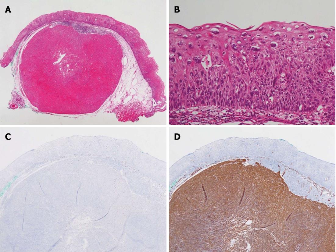
Figure 3 Histopathologic findings.
A: Esophageal leiomyoma arising from the muscularis mucosa (HE, 20 ×); B: Atypical cells in all three areas of the epithelium, but not full thickness, showed severe dysplasia (HE, 200 ×); C: The esophageal leiomyoma was negative for CD117 (C-Kit); D: The esophageal leiomyoma showed strong and diffuse positive staining for smooth muscle actin (200 ×).
- Citation: Ahn SY, Jeon SW. Endoscopic resection of co-existing severe dysplasia and a small esophageal leiomyoma. World J Gastroenterol 2013; 19(1): 137-140
- URL: https://www.wjgnet.com/1007-9327/full/v19/i1/137.htm
- DOI: https://dx.doi.org/10.3748/wjg.v19.i1.137


