Copyright
©2013 Baishideng Publishing Group Co.
World J Gastroenterol. Jan 7, 2013; 19(1): 137-140
Published online Jan 7, 2013. doi: 10.3748/wjg.v19.i1.137
Published online Jan 7, 2013. doi: 10.3748/wjg.v19.i1.137
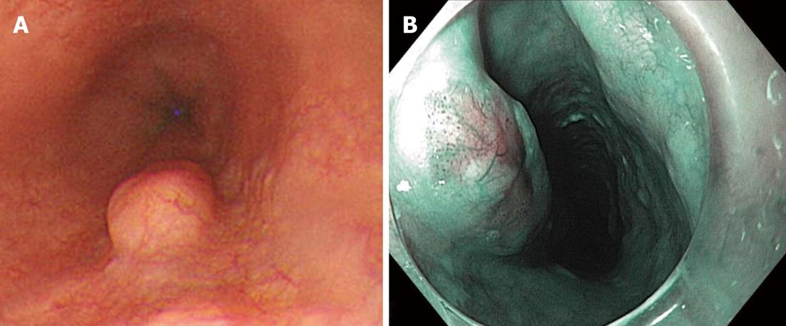
Figure 1 Endoscopic findings.
A: Endoscopic findings demonstrate a subepithelial tumor with intact overlying mucosa; B: Chromoendoscopy with narrow band imaging showed scattered brown dots, and dilated and tortuous vessels on the top of the lesion.
- Citation: Ahn SY, Jeon SW. Endoscopic resection of co-existing severe dysplasia and a small esophageal leiomyoma. World J Gastroenterol 2013; 19(1): 137-140
- URL: https://www.wjgnet.com/1007-9327/full/v19/i1/137.htm
- DOI: https://dx.doi.org/10.3748/wjg.v19.i1.137


