Copyright
©2013 Baishideng Publishing Group Co.
World J Gastroenterol. Jan 7, 2013; 19(1): 133-136
Published online Jan 7, 2013. doi: 10.3748/wjg.v19.i1.133
Published online Jan 7, 2013. doi: 10.3748/wjg.v19.i1.133
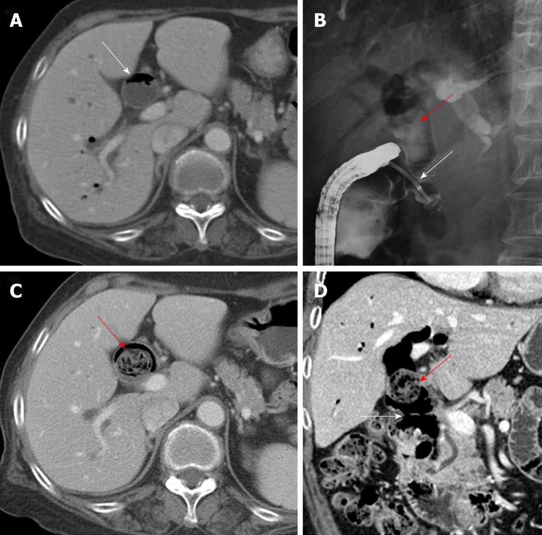
Figure 1 Development of a biliary phytobezoar in the extrahepatic bile duct.
A: Axial portal venous-phase computed tomography (PVP CT) 3 years ago showing diffuse bile duct dilatation (white arrow) with pneumobilia; B: On endoscopic retrograde cholangiopancreatography, there was no abnormal filling defect except pneumobilia (red arrow) in the dilated extrahepatic bile duct (EHD). A choledochoduodenal fistula (white arrow) in the distal common bile duct which was cannulated by endoscopy; C and D: Axial PVP CT and its coronal reformatted image at 6-mo follow-up showed a dilated EHD and a newly developed intraductal ovoid mass with interstitial air (red arrows), as well as the choledochoduodenalfistula (white arrow).
- Citation: Kim Y, Park BJ, Kim MJ, Sung DJ, Kim DS, Yu YD, Lee JH. Biliary phytobezoar resulting in intestinal obstruction. World J Gastroenterol 2013; 19(1): 133-136
- URL: https://www.wjgnet.com/1007-9327/full/v19/i1/133.htm
- DOI: https://dx.doi.org/10.3748/wjg.v19.i1.133


