Copyright
©2013 Baishideng Publishing Group Co.
World J Gastroenterol. Jan 7, 2013; 19(1): 129-132
Published online Jan 7, 2013. doi: 10.3748/wjg.v19.i1.129
Published online Jan 7, 2013. doi: 10.3748/wjg.v19.i1.129
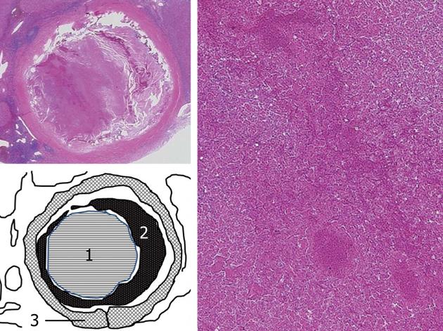
Figure 3 Schematic image of the structure of the calcified lesion.
Upper left, loupe image of the calcified lesion (hematoxylin and eosin staining; original magnification, × 5); Right, microscopic findings of the calcified lesion showed that necrotic area was extensively observed in the tumor (hematoxylin and eosin staining; original magnification, × 40); Lower left, schematic of the structure of the calcified lesion. A poorly differentiated tumor, in which spotty necrosis and hemorrhagic necrosis were extensively observed, was surrounded by a fibrous capsule. Calcification was identified between the tumor and the fibrous capsule (1: Poorly differentiated tumor; 2: Calcification; 3: Fibrous capsule).
- Citation: Murakami T, Morioka D, Takakura H, Miura Y, Togo S. Small hepatocellular carcinoma with ring calcification: A case report and literature review. World J Gastroenterol 2013; 19(1): 129-132
- URL: https://www.wjgnet.com/1007-9327/full/v19/i1/129.htm
- DOI: https://dx.doi.org/10.3748/wjg.v19.i1.129


