Copyright
©2013 Baishideng Publishing Group Co.
World J Gastroenterol. Jan 7, 2013; 19(1): 125-128
Published online Jan 7, 2013. doi: 10.3748/wjg.v19.i1.125
Published online Jan 7, 2013. doi: 10.3748/wjg.v19.i1.125
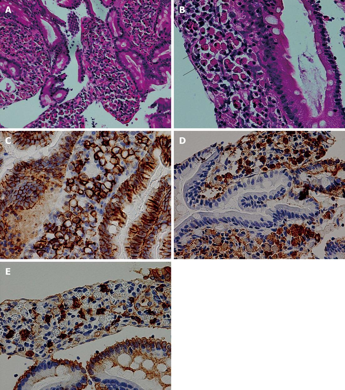
Figure 3 Histological appearance of the duodenal biopsy.
A, B: Numerous Mott cells (arrows), whose cytoplasm is filled with eosinophilic inclusion bodies (Russell bodies), infiltrate the lamina propria mucosae (HE stain, A: × 200, B: × 400); C: Immunohistochemically, Mott cells are positive for CD138 (× 400); D, E: Infiltrating plasma cells show a polyclonal pattern upon the immunohistochemical staining for κ (D) and λ (E) chains (× 400).
- Citation: Takahashi Y, Shimizu S, Uraushihara K, Fukusato T. Russell body duodenitis in a patient with retroperitoneal metastasis of ureteral cancer. World J Gastroenterol 2013; 19(1): 125-128
- URL: https://www.wjgnet.com/1007-9327/full/v19/i1/125.htm
- DOI: https://dx.doi.org/10.3748/wjg.v19.i1.125


