Copyright
©2012 Baishideng Publishing Group Co.
World J Gastroenterol. Feb 21, 2012; 18(7): 704-711
Published online Feb 21, 2012. doi: 10.3748/wjg.v18.i7.704
Published online Feb 21, 2012. doi: 10.3748/wjg.v18.i7.704
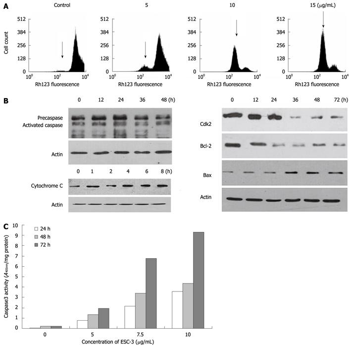
Figure 4 Apoptosis induced by ESC-3 through the mitochondria-dependent pathway.
A: Effect of ESC-3 on the ΔΨm of cholangiocarcinoma cells. The increase in Rh123 hypofluorescence indicates a reduction in ΔΨm, which is shown with arrows; B: Expression of cytochrome C, caspase-3, CDK2, Bax, and Bcl-2 in Mz-ChA-1 cells treated with 10 μg/mL ESC-3 for different periods of time; C: Effect of ESC-3 on the activation of caspase-3 activity in Mz-ChA-1 cells. The cells were treated with 0 μg/mL, 5 μg/mL, 7.5 μg/mL and 12.5 μg/mL ESC-3, and caspase-3 activity was analyzed after 24 h, 48 h or 72 h.
-
Citation: Song W, Shen DY, Kang JH, Li SS, Zhan HW, Shi Y, Xiong YX, Liang G, Chen QX. Apoptosis of human cholangiocarcinoma cells induced by ESC-3 from
Crocodylus siamensis bile. World J Gastroenterol 2012; 18(7): 704-711 - URL: https://www.wjgnet.com/1007-9327/full/v18/i7/704.htm
- DOI: https://dx.doi.org/10.3748/wjg.v18.i7.704


