Copyright
©2012 Baishideng Publishing Group Co.
World J Gastroenterol. Feb 14, 2012; 18(6): 507-516
Published online Feb 14, 2012. doi: 10.3748/wjg.v18.i6.507
Published online Feb 14, 2012. doi: 10.3748/wjg.v18.i6.507
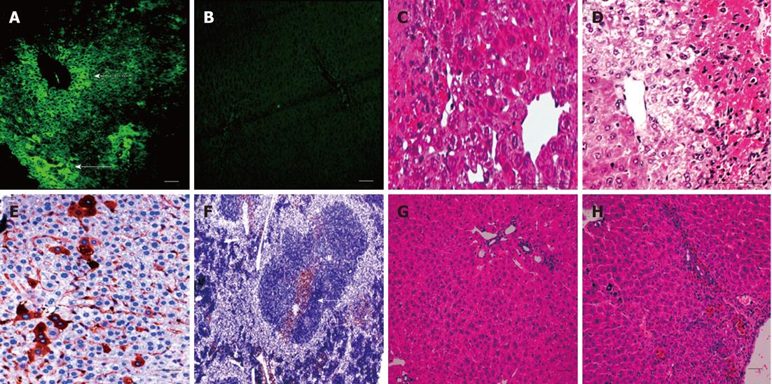
Figure 4 Embryonic stem cell liver engraftment following acetaminophen induced damage.
A and B: Green fluorescence protein (GFP) +ve cells were present under the liver capsule (a-solid arrow) and around the central veins of the liver at 72 h, as seen under direct fluorescence (a-dashed arrow) with no fluorescence detected in the control group (B); C and D: The characteristic pattern of acetaminophen-induced liver damage after 72 h mainly affected the centrilobular portions of the liver, with marked damage of the pericentral hepatocytes in both the cell therapy and control groups, although pericentral vacuolation was more evident in the control group; E: At 2 wk, the GFP +ve cells could be detected using IHC in the hepatic parenchyma and within the sinusoidal lining; F: Localized colonies of GFP +ve cells were also detected in the spleen at 2 wk; G and H: After 2 wk the liver recovered in both groups, with the liver sections from the cell therapy group (G) showing less inflammatory cells and congestion than in the control group (H) (scale bar = 200 μ).
- Citation: Ezzat T, Dhar DK, Malago M, Damink SWO. Dynamic tracking of stem cells in an acute liver failure model. World J Gastroenterol 2012; 18(6): 507-516
- URL: https://www.wjgnet.com/1007-9327/full/v18/i6/507.htm
- DOI: https://dx.doi.org/10.3748/wjg.v18.i6.507


