Copyright
©2012 Baishideng Publishing Group Co.
World J Gastroenterol. Feb 14, 2012; 18(6): 507-516
Published online Feb 14, 2012. doi: 10.3748/wjg.v18.i6.507
Published online Feb 14, 2012. doi: 10.3748/wjg.v18.i6.507
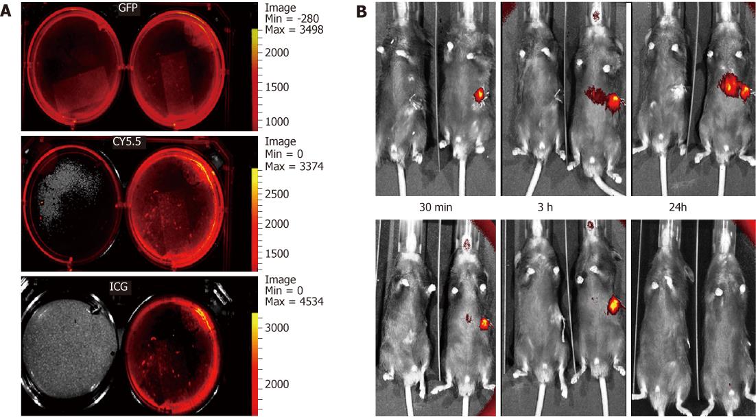
Figure 2 Labeling and tracking of the fluorescent embryonic stem cell following acetaminophen administration.
IVIS images of green fluorescence protein (GFP) +ve cultured embryonic stem cells (ESCs) without 1,1-dioctadecyl-3,3,3,3-tetramethylindotricarbocyanine iodide (DiR) staining (LT) and with DiR staining (RT) showing background fluorescence using the GFP and tricarbocyanine 5.5 filters, but not with the indocyanine green (ICG) filter (A). Images of a pair of mice (B), one from the cell therapy group-RT and the other from the control group-LT, were compared using IVIS. At 30 min following transplantation, a strong signal could only be detected from the spleen where the cells were injected. Between 3 and 24 h following the cell transplantation, the signal started to intensify between the spleen and the liver, which is most probably the splenic vein owing to its tortuous course. A strong signal was detected in the liver over 24 h post-transplantation, which faded out by the 72 h time-period. After one week, the signal could not be detected in the liver, but was still strong and was detectable over the spleen, which had completely disappeared by 2 wk.
- Citation: Ezzat T, Dhar DK, Malago M, Damink SWO. Dynamic tracking of stem cells in an acute liver failure model. World J Gastroenterol 2012; 18(6): 507-516
- URL: https://www.wjgnet.com/1007-9327/full/v18/i6/507.htm
- DOI: https://dx.doi.org/10.3748/wjg.v18.i6.507


