Copyright
©2012 Baishideng Publishing Group Co.
World J Gastroenterol. Feb 7, 2012; 18(5): 472-478
Published online Feb 7, 2012. doi: 10.3748/wjg.v18.i5.472
Published online Feb 7, 2012. doi: 10.3748/wjg.v18.i5.472
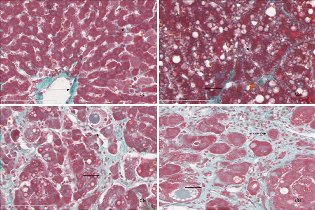
Figure 2 Collagen distributions in rat model livers (Masson × 40).
Normal hepatic structure (d0) was demonstrated in control rats administered 3 mL/kg olive oil subcutaneously twice weekly for 84 d. Hepatic degeneration (d28), fibrosis (d56) and cirrhosis (d84) were seen in the chronic hepatitis model rats administered 3 mL/kg 40% (v/v) CCl4 in olive oil subcutaneously twice weekly for 28 d, 56 d, and 84 d, respectively. There was less collagen mainly around the central veins (solid arrow) and the portal areas, with some staining noise signals in lobules (dashed arrow) in normal hepatic structure (d0). The collagen fiber bundles (solid arrow) had partly spread into the lobules from the portal areas, the hepatic cells had obvious watery and fatty degeneration (dashed arrow) and had not been isolated by collagen during hepatic degeneration (d28). The collagen fiber bundles (solid arrow) had completely separated some of the lobules to form many pseudo-lobules, several hepatic cells with obvious fatty degeneration (dashed arrow) had been completely isolated by collagen in hepatic fibrosis (d56). The collagen fiber bundles (solid arrow) had limited smaller pseudo-lobules, some single hepatic cells (dashed arrow) were completely isolated by collagen in hepatic cirrhosis (d84).
- Citation: Zhang T, Xu XY, Zhou H, Zhao X, Song M, Zhang TT, Yin H, Li T, Li PT, Cai DY. A pharmacodynamic model of portal hypertension in isolated perfused rat liver. World J Gastroenterol 2012; 18(5): 472-478
- URL: https://www.wjgnet.com/1007-9327/full/v18/i5/472.htm
- DOI: https://dx.doi.org/10.3748/wjg.v18.i5.472


