Copyright
©2012 Baishideng Publishing Group Co.
World J Gastroenterol. Feb 7, 2012; 18(5): 435-444
Published online Feb 7, 2012. doi: 10.3748/wjg.v18.i5.435
Published online Feb 7, 2012. doi: 10.3748/wjg.v18.i5.435
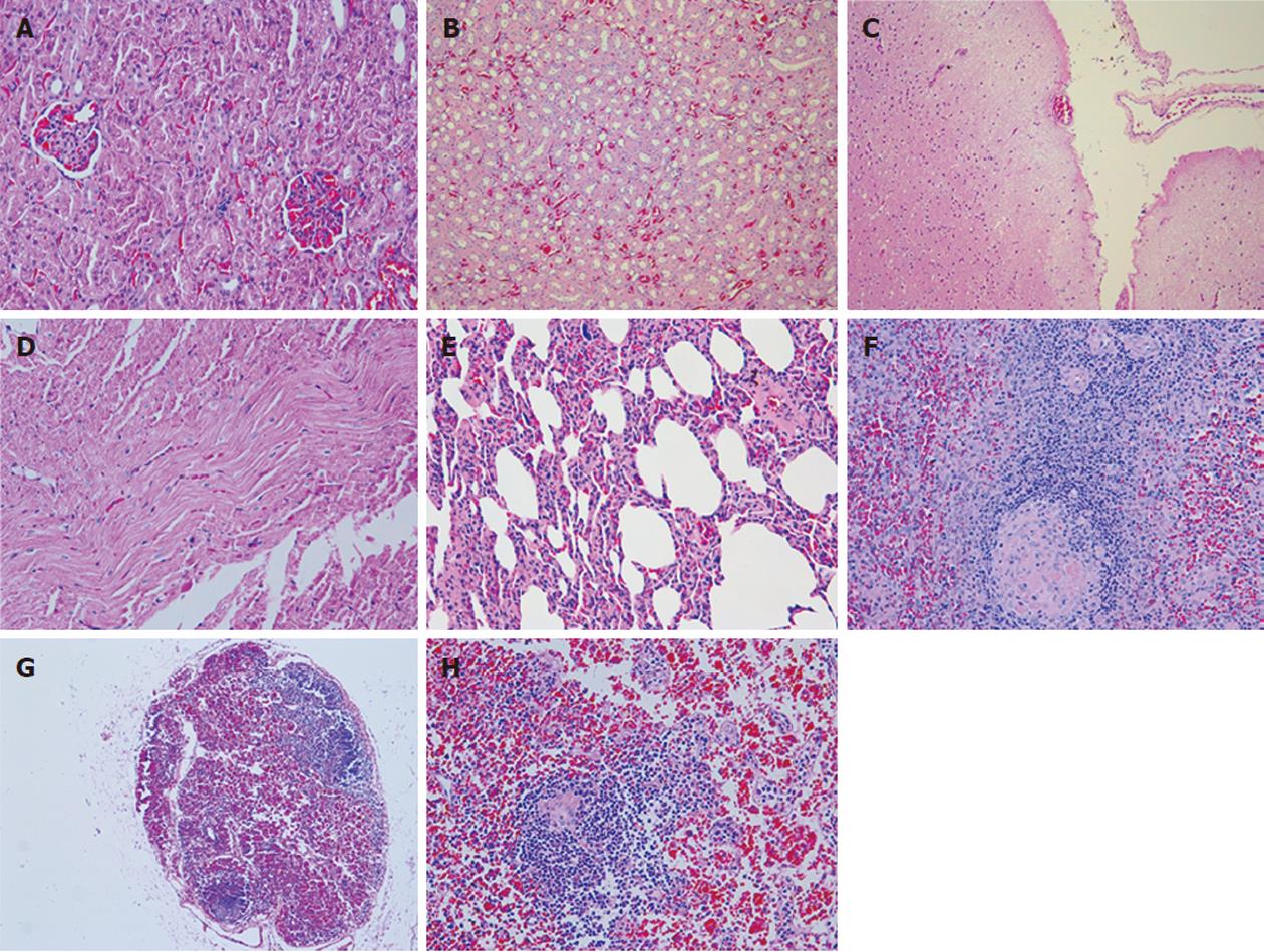
Figure 6 Histopathological changes of other organs.
A: Ectasia and hyperemia of capillaries in the renal glomerulus [hematoxylin and eosin (H and E) stain, × 200]; B: Widespread hyperemia of capillaries in the renal stroma and cellular swelling in the tubular epithelial cells (H and E stain, × 100); C: Weak staining in the superficial layer of the brain suggest cerebral edema (H and E stain, × 100); D: Wave-like changes in the myocardium (H and E stain, × 200); E: Hyperemia of capillaries in the stroma and hyaline changes in the arterioles of the lung (H and E stain, × 200); F: Hyaline changes in the central artery of the spleen (H and E stain, × 200); G: Widespread reduction in lymphocytes and hemorrhage in the mesenteric lymph nodes (H and E stain, × 50); H: Hemorrhage and histiocyte proliferation in the mesenteric lymph nodes. Hyaline change appeared in the vessel wall of lymphoid follicles (H and E stain, × 200).
-
Citation: Zhou P, Xia J, Guo G, Huang ZX, Lu Q, Li L, Li HX, Shi YJ, Bu H. A
Macaca mulatta model of fulminant hepatic failure. World J Gastroenterol 2012; 18(5): 435-444 - URL: https://www.wjgnet.com/1007-9327/full/v18/i5/435.htm
- DOI: https://dx.doi.org/10.3748/wjg.v18.i5.435


