Copyright
©2012 Baishideng Publishing Group Co.
World J Gastroenterol. Dec 28, 2012; 18(48): 7397-7401
Published online Dec 28, 2012. doi: 10.3748/wjg.v18.i48.7397
Published online Dec 28, 2012. doi: 10.3748/wjg.v18.i48.7397
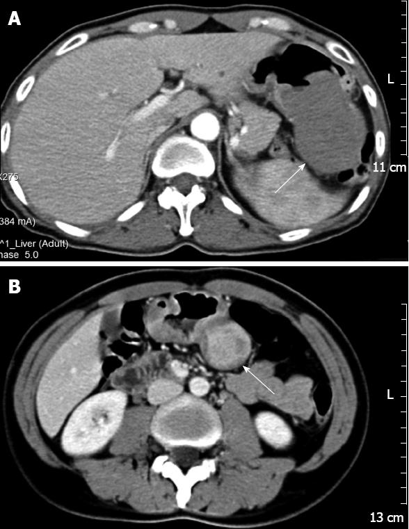
Figure 1 Computed tomography images showed rounded masses in the stomachs, with homogeneous (n = 3) or heterogeneous (n = 1) internal contrast enhancement.
A: Contrast-enhanced computed tomography (CT) showed a solitary, exophytic, soft, internal homogeneous tissue mass (arrow) in the greater curvature of the stomach, the mass exhibited central ulceration (Case 3); B: CT during the portal venous phase of contrast enhancement showed a heterogeneous contrast enhanced mass (arrow) in the body of the stomach (Case 1).
- Citation: Zhong DD, Wang CH, Xu JH, Chen MY, Cai JT. Endoscopic ultrasound features of gastric schwannomas with radiological correlation: A case series report. World J Gastroenterol 2012; 18(48): 7397-7401
- URL: https://www.wjgnet.com/1007-9327/full/v18/i48/7397.htm
- DOI: https://dx.doi.org/10.3748/wjg.v18.i48.7397


