Copyright
©2012 Baishideng Publishing Group Co.
World J Gastroenterol. Dec 21, 2012; 18(47): 7104-7108
Published online Dec 21, 2012. doi: 10.3748/wjg.v18.i47.7104
Published online Dec 21, 2012. doi: 10.3748/wjg.v18.i47.7104
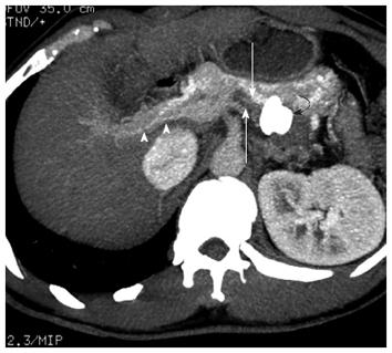
Figure 4 Axial image of contrast enhanced 64-slice helical computed tomography scan of the abdomen, showing the correct Amplatzer vascular plug position (curved arrow), opacification of the portal system (arrow heads), and a thrombosed shunt tract (arrows).
- Citation: Wang MQ, Liu FY, Duan F. Management of surgical splenorenal shunt-related hepatic myelopathy with endovascular interventional techniques. World J Gastroenterol 2012; 18(47): 7104-7108
- URL: https://www.wjgnet.com/1007-9327/full/v18/i47/7104.htm
- DOI: https://dx.doi.org/10.3748/wjg.v18.i47.7104


