Copyright
©2012 Baishideng Publishing Group Co.
World J Gastroenterol. Dec 21, 2012; 18(47): 7104-7108
Published online Dec 21, 2012. doi: 10.3748/wjg.v18.i47.7104
Published online Dec 21, 2012. doi: 10.3748/wjg.v18.i47.7104
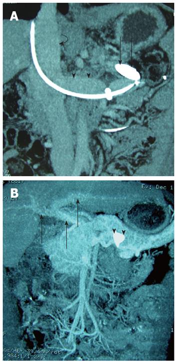
Figure 2 Coronal maximum intensity projection reconstruction of a contrast enhanced 64-slice helical computed tomography scan of the abdomen.
A: Obtained 5 d after placement of an occlusion through the splenorenal shunt and shows the occlusion balloon placed within the shunt tract (arrows), note the inferior vena cava (curved arrow) and the left renal vein (arrow heads); B: Obtained 3 mo after the Amplatzer vascular plugs (AVP) occlusion procedure and shows the correct AVP position (arrow heads), opacification of the portal system (arrows), and no opacification of the surgical shunt, as well as the varices.
- Citation: Wang MQ, Liu FY, Duan F. Management of surgical splenorenal shunt-related hepatic myelopathy with endovascular interventional techniques. World J Gastroenterol 2012; 18(47): 7104-7108
- URL: https://www.wjgnet.com/1007-9327/full/v18/i47/7104.htm
- DOI: https://dx.doi.org/10.3748/wjg.v18.i47.7104


