Copyright
©2012 Baishideng Publishing Group Co.
World J Gastroenterol. Dec 21, 2012; 18(47): 7070-7078
Published online Dec 21, 2012. doi: 10.3748/wjg.v18.i47.7070
Published online Dec 21, 2012. doi: 10.3748/wjg.v18.i47.7070
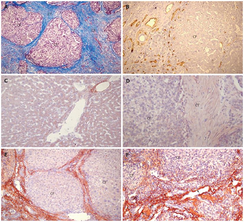
Figure 1 Expression and distribution characteristics of transforming growth factor β1 or connective tissue growth factor mRNA in human hepatocellular carcinoma.
A: Masson’s trichrome stain for collagen (blue) in hepatocellular carcinoma (HCC); B: Immunohistochemical detection of CD34 in vascular endothelial cells (brown) in HCC; C: Normal liver showing connective tissue growth factor (CCN2) mRNA expressed in connective tissue around the veins; D: Absence of staining of HCC when the in situ hybridization probes were omitted; E: HCC stained for transforming growth factor β1 mRNA, showing reactivity in connective tissue and around the carcinoma foci; F: Over-expression of CCN2 mRNA in connective tissue and vascular endothelial cells (black arrow) in HCC. Original magnification, × 100 in A and E, × 200 in B, C, D and F. CF: Carcinoma foci; CT: Connective tissue.
- Citation: Xiu M, Liu YH, Brigstock DR, He FH, Zhang RJ, Gao RP. Connective tissue growth factor is overexpressed in human hepatocellular carcinoma and promotes cell invasion and growth. World J Gastroenterol 2012; 18(47): 7070-7078
- URL: https://www.wjgnet.com/1007-9327/full/v18/i47/7070.htm
- DOI: https://dx.doi.org/10.3748/wjg.v18.i47.7070


