Copyright
©2012 Baishideng Publishing Group Co.
World J Gastroenterol. Dec 21, 2012; 18(47): 7048-7055
Published online Dec 21, 2012. doi: 10.3748/wjg.v18.i47.7048
Published online Dec 21, 2012. doi: 10.3748/wjg.v18.i47.7048
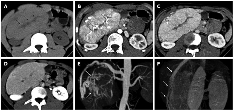
Figure 2 Focal nodular hyperplasia in a 16-year-old boy.
A: The lesion was hypodense on pre-contrast computed tomography scan, and central scar with much lower density were identified (arrows); B: The lesion was significantly enhanced in the arterial phase with enlarged feeding arteries penetrating into the central scar (arrows); C: The lesion was slightly hyperdense in the portal vein phase; D: The lesion was isodense in the equilibrium phase with delayed enhancement of the central scar; E: Computed tomography angiography (CTA) in the arterial phase showed that the enlarged feeding arteries were distorted, and exhibited a spoke-wheel shaped blood supply (arrows); F: CTA in the portal vein phase showed the draining hepatic vein (arrows).
- Citation: Liu QY, Zhang WD, Lai DM, Ou-yang Y, Gao M, Lin XF. Hepatic focal nodular hyperplasia in children: Imaging features on multi-slice computed tomography. World J Gastroenterol 2012; 18(47): 7048-7055
- URL: https://www.wjgnet.com/1007-9327/full/v18/i47/7048.htm
- DOI: https://dx.doi.org/10.3748/wjg.v18.i47.7048


