Copyright
©2012 Baishideng Publishing Group Co.
World J Gastroenterol. Dec 21, 2012; 18(47): 6974-6980
Published online Dec 21, 2012. doi: 10.3748/wjg.v18.i47.6974
Published online Dec 21, 2012. doi: 10.3748/wjg.v18.i47.6974
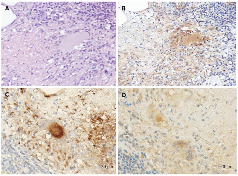
Figure 2 Histopathological views of tuberculous granuloma and localization of the mycobacterial antigen in the colonic lesion (patient 8).
A: A granuloma, surrounded by inflammatory lymphocytes, is present in the lamina propria (hematoxylin and eosin, × 200); B: Immunohistochemical staining view of the colonic specimen (× 200). Note that the mycobacterial antigens (brownish granular matter) are present in the cytoplasm of epithelioid histiocytes and multinucleated giant cells in the granuloma; C: Cells stained with the pan-macrophage marker CD68 antibody are present in the granuloma (× 400); D: The mycobacterial antigens (brownish granular matter) are present in the cytoplasm of the macrophages in the granuloma (× 400).
-
Citation: Ihama Y, Hokama A, Hibiya K, Kishimoto K, Nakamoto M, Hirata T, Kinjo N, Cash HL, Higa F, Tateyama M, Kinjo F, Fujita J. Diagnosis of intestinal tuberculosis using a monoclonal antibody to
Mycobacterium tuberculosis . World J Gastroenterol 2012; 18(47): 6974-6980 - URL: https://www.wjgnet.com/1007-9327/full/v18/i47/6974.htm
- DOI: https://dx.doi.org/10.3748/wjg.v18.i47.6974


