Copyright
©2012 Baishideng Publishing Group Co.
World J Gastroenterol. Dec 14, 2012; 18(46): 6790-6800
Published online Dec 14, 2012. doi: 10.3748/wjg.v18.i46.6790
Published online Dec 14, 2012. doi: 10.3748/wjg.v18.i46.6790
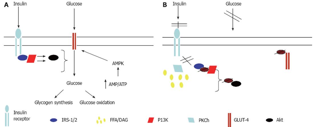
Figure 2 Mechanisms for free fatty acid-induced skeletal muscle insulin resistance.
A: Normal glucose uptake into skeletal muscle occurs via binding of insulin to the insulin receptor, resulting in receptor autophosphorylation and subsequent binding and tyrosine phosphorylation of insulin receptor substrate (IRS)-1. Subsequently, phosphatidylinositol 3-kinase (PI3K) is activated and results in downstream activation of protein kinase B, leading to glucose transporter (GLUT)-4 translocation to the myocyte plasma membrane and glucose uptake into the cell; B: Increased intramyocellular lipid content leads to the activation of protein kinase Ch (PKCh) which results in serine phosphorylation of IRS-1, thereby inhibiting its tyrosine phosphorylation. This prevents the activation of PI3K and GLUT-4 translocation to the cell surface. Thus glucose entry into the cell is inhibited. AMPK: AMP-activated protein kinase; DAG: Diacylglycerol; FFA: Free fatty acid; Akt: Protein Kinase B.
- Citation: Finelli C, Tarantino G. Have guidelines addressing physical activity been established in nonalcoholic fatty liver disease? World J Gastroenterol 2012; 18(46): 6790-6800
- URL: https://www.wjgnet.com/1007-9327/full/v18/i46/6790.htm
- DOI: https://dx.doi.org/10.3748/wjg.v18.i46.6790


