Copyright
©2012 Baishideng Publishing Group Co.
World J Gastroenterol. Dec 7, 2012; 18(45): 6677-6681
Published online Dec 7, 2012. doi: 10.3748/wjg.v18.i45.6677
Published online Dec 7, 2012. doi: 10.3748/wjg.v18.i45.6677
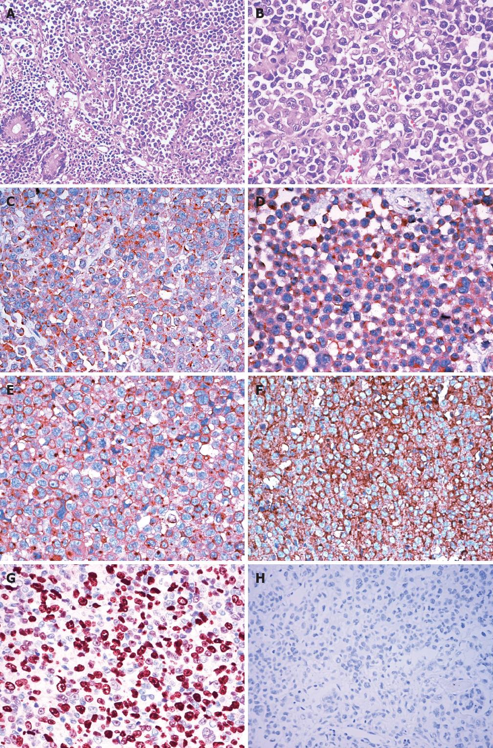
Figure 1 Plasmablastic lymphoma of the small intestine.
A: Diffuse infiltration in the mucosa of the small intestinal by monotonous large atypical lymphoid cells [hematoxylin and eosin (H and E), original magnification ×200]; B: These atypical cells had a plasmablastic appearance, with abundant basophilic cytoplasm, eccentrically located pleomorphic nuclei, and single, centrally located prominent nucleoli (H and E, original magnification ×400); C-F: The atypical cells were diffusely positive for CD79a (C), CD138 (D), CD10 (E) and immunoglobulin light chain κ (F) (immunoperoxidase stain, original magnification ×400); G: Nuclear proliferation rate as assessed by Ki-67 staining was approximately 80% (immunoperoxidase stain, original magnification ×400); H: Epstein-Barr virus (EBV)-encoded RNA in situ hybridization for EBV shows negative staining in the nucleus of these atypical cells (original magnification ×400).
- Citation: Wang HW, Yang W, Sun JZ, Lu JY, Li M, Sun L. Plasmablastic lymphoma of the small intestine: Case report and literature review. World J Gastroenterol 2012; 18(45): 6677-6681
- URL: https://www.wjgnet.com/1007-9327/full/v18/i45/6677.htm
- DOI: https://dx.doi.org/10.3748/wjg.v18.i45.6677


