Copyright
©2012 Baishideng Publishing Group Co.
World J Gastroenterol. Nov 28, 2012; 18(44): 6504-6509
Published online Nov 28, 2012. doi: 10.3748/wjg.v18.i44.6504
Published online Nov 28, 2012. doi: 10.3748/wjg.v18.i44.6504
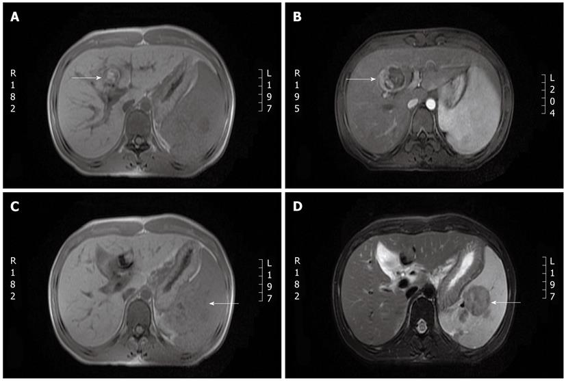
Figure 3 Liver magnetic resonance imaging.
A: A 2.3 cm × 2.56 cm lesion was observed in segment 4 of the liver (T1-weighted). No obvious dilation of the intra or extrahepatic bile duct was observed; B: Following contrast magnetic resonance imaging (MRI), the capsule shows linear enhancement without enhancement of the lesion itself. Splenomegaly is observed, and several oval-shaped nodules in the spleen show slight enhancement; C: The oval-shaped nodules appear as low-intensity signals on a T1-weighted MRI image; D: The oval-shaped nodules in the spleen appear as a lower-intensity signal on T2-weighted MRI images than on T1-weighted images.
-
Citation: Deng BC, Lv S, Cui W, Zhao R, Lu X, Wu J, Liu P. Novel
ATP8B1 mutation in an adult male with progressive familial intrahepatic cholestasis. World J Gastroenterol 2012; 18(44): 6504-6509 - URL: https://www.wjgnet.com/1007-9327/full/v18/i44/6504.htm
- DOI: https://dx.doi.org/10.3748/wjg.v18.i44.6504


