Copyright
©2012 Baishideng Publishing Group Co.
World J Gastroenterol. Nov 28, 2012; 18(44): 6468-6474
Published online Nov 28, 2012. doi: 10.3748/wjg.v18.i44.6468
Published online Nov 28, 2012. doi: 10.3748/wjg.v18.i44.6468
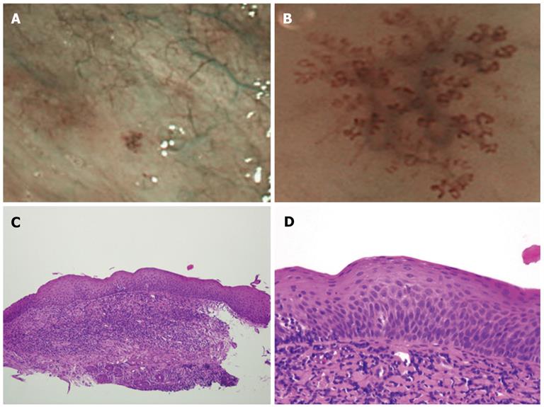
Figure 2 Narrow-band imaging view and histological image of low-grade dysplasia.
A: Narrow-band imaging (NBI) showing the oropharynx with a well-demarcated brownish area; B: A magnified NBI magnifying view showing an intra-papillary capillary loop type IV pattern; C: Low power magnification of the biopsied specimen [hematoxylin and eosin (HE), original ×100]; D: Histologically, the lesion showed minimal cell size variation and was diagnosed as low-grade dysplasia (HE, original × 400).
- Citation: Kumamoto T, Sentani K, Oka S, Tanaka S, Yasui W. Clinicopathological features of minute pharyngeal lesions diagnosed by narrow-band imaging endoscopy and biopsy. World J Gastroenterol 2012; 18(44): 6468-6474
- URL: https://www.wjgnet.com/1007-9327/full/v18/i44/6468.htm
- DOI: https://dx.doi.org/10.3748/wjg.v18.i44.6468


