Copyright
©2012 Baishideng Publishing Group Co.
World J Gastroenterol. Nov 28, 2012; 18(44): 6427-6436
Published online Nov 28, 2012. doi: 10.3748/wjg.v18.i44.6427
Published online Nov 28, 2012. doi: 10.3748/wjg.v18.i44.6427
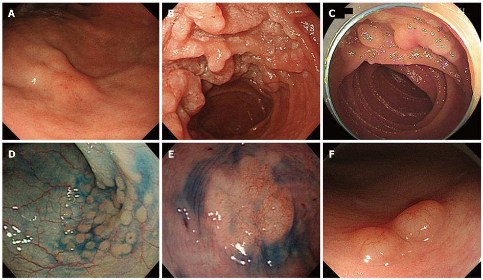
Figure 2 Endoscopic images of follicular lymphoma.
A: A gastric lesion with thickened rugae exhibiting a slight redness; B: Typical features of the whitish polypoid granules observed in the duodenum; C: Whitish polypoid lesions in the jejunum; D: Indigo carmine contrast was used to emphasize the slightly elevated small polyps present in the colon; E: An elevated lesion with a flat surface and a 20 mm diameter was observed in the rectum; F: In another patient, polypoid lesions exhibiting hypervascularity on the surface were detected in the rectum.
- Citation: Iwamuro M, Okada H, Takata K, Shinagawa K, Fujiki S, Shiode J, Imagawa A, Araki M, Morito T, Nishimura M, Mizuno M, Inaba T, Suzuki S, Kawai Y, Yoshino T, Kawahara Y, Takaki A, Yamamoto K. Diagnostic role of 18F-fluorodeoxyglucose positron emission tomography for follicular lymphoma with gastrointestinal involvement. World J Gastroenterol 2012; 18(44): 6427-6436
- URL: https://www.wjgnet.com/1007-9327/full/v18/i44/6427.htm
- DOI: https://dx.doi.org/10.3748/wjg.v18.i44.6427


