Copyright
©2012 Baishideng Publishing Group Co.
World J Gastroenterol. Nov 28, 2012; 18(44): 6427-6436
Published online Nov 28, 2012. doi: 10.3748/wjg.v18.i44.6427
Published online Nov 28, 2012. doi: 10.3748/wjg.v18.i44.6427
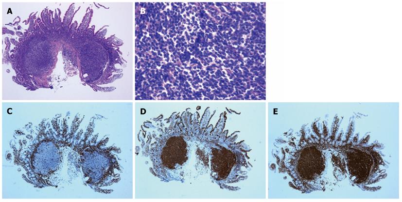
Figure 1 Typical histological features of follicular lymphoma.
A, B: Small cleaved cells that infiltrated the duodenal mucosa and formed lymphoid follicles are present (hematoxylin and eosin staining); C: Representative immunohistochemical staining of lymphoma cells negative for CD3; D: Lymphoma cells positive for CD10; E: Lymphoma cells positive for BCL-2. All of the images shown are at 40 × magnification, except for panel B which is at 400 × magnification.
- Citation: Iwamuro M, Okada H, Takata K, Shinagawa K, Fujiki S, Shiode J, Imagawa A, Araki M, Morito T, Nishimura M, Mizuno M, Inaba T, Suzuki S, Kawai Y, Yoshino T, Kawahara Y, Takaki A, Yamamoto K. Diagnostic role of 18F-fluorodeoxyglucose positron emission tomography for follicular lymphoma with gastrointestinal involvement. World J Gastroenterol 2012; 18(44): 6427-6436
- URL: https://www.wjgnet.com/1007-9327/full/v18/i44/6427.htm
- DOI: https://dx.doi.org/10.3748/wjg.v18.i44.6427


