Copyright
©2012 Baishideng Publishing Group Co.
World J Gastroenterol. Nov 21, 2012; 18(43): 6341-6344
Published online Nov 21, 2012. doi: 10.3748/wjg.v18.i43.6341
Published online Nov 21, 2012. doi: 10.3748/wjg.v18.i43.6341
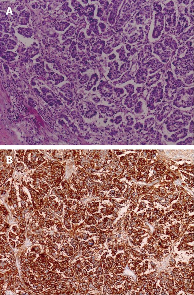
Figure 4 Pathological manifestation of the neoplasm.
A: Microscopcally, the tumor is composed of irregular sheets of cells with a high-grade nuclear atypia (HE stain, × 100); B: Immunohistochemically, the tumor cells are immunoreactive for cancer antigen 125 (× 100).
- Citation: Zhou JJ, Miao XY. Gastric metastasis from ovarian carcinoma: A case report and literature review. World J Gastroenterol 2012; 18(43): 6341-6344
- URL: https://www.wjgnet.com/1007-9327/full/v18/i43/6341.htm
- DOI: https://dx.doi.org/10.3748/wjg.v18.i43.6341


