Copyright
©2012 Baishideng Publishing Group Co.
World J Gastroenterol. Nov 21, 2012; 18(43): 6338-6340
Published online Nov 21, 2012. doi: 10.3748/wjg.v18.i43.6338
Published online Nov 21, 2012. doi: 10.3748/wjg.v18.i43.6338
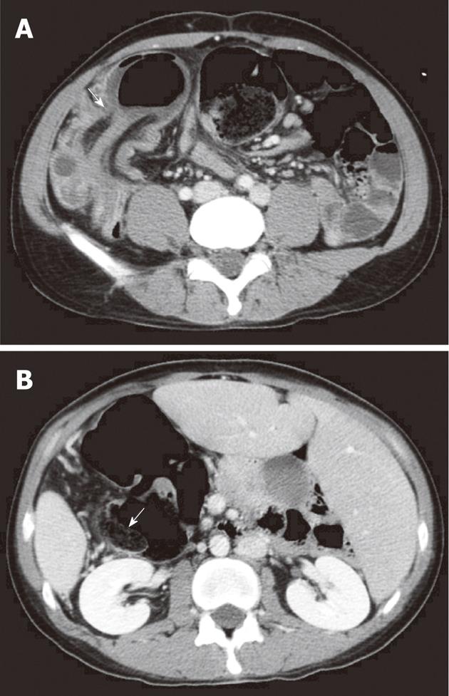
Figure 2 Axial computed tomographic scans of the lower abdomen.
A: Intussusception in the region of the sigmoid and descending colon at right lower quadrant of the abdomen (arrow); B: Scan at a more cranial level revealing a distended descending colon with a thin enhancing rim of a septum (arrow).
- Citation: Ho YC. Total colorectal and terminal ileal duplication presenting as intussusception and intestinal obstruction. World J Gastroenterol 2012; 18(43): 6338-6340
- URL: https://www.wjgnet.com/1007-9327/full/v18/i43/6338.htm
- DOI: https://dx.doi.org/10.3748/wjg.v18.i43.6338


