Copyright
©2012 Baishideng Publishing Group Co.
World J Gastroenterol. Nov 21, 2012; 18(43): 6328-6332
Published online Nov 21, 2012. doi: 10.3748/wjg.v18.i43.6328
Published online Nov 21, 2012. doi: 10.3748/wjg.v18.i43.6328
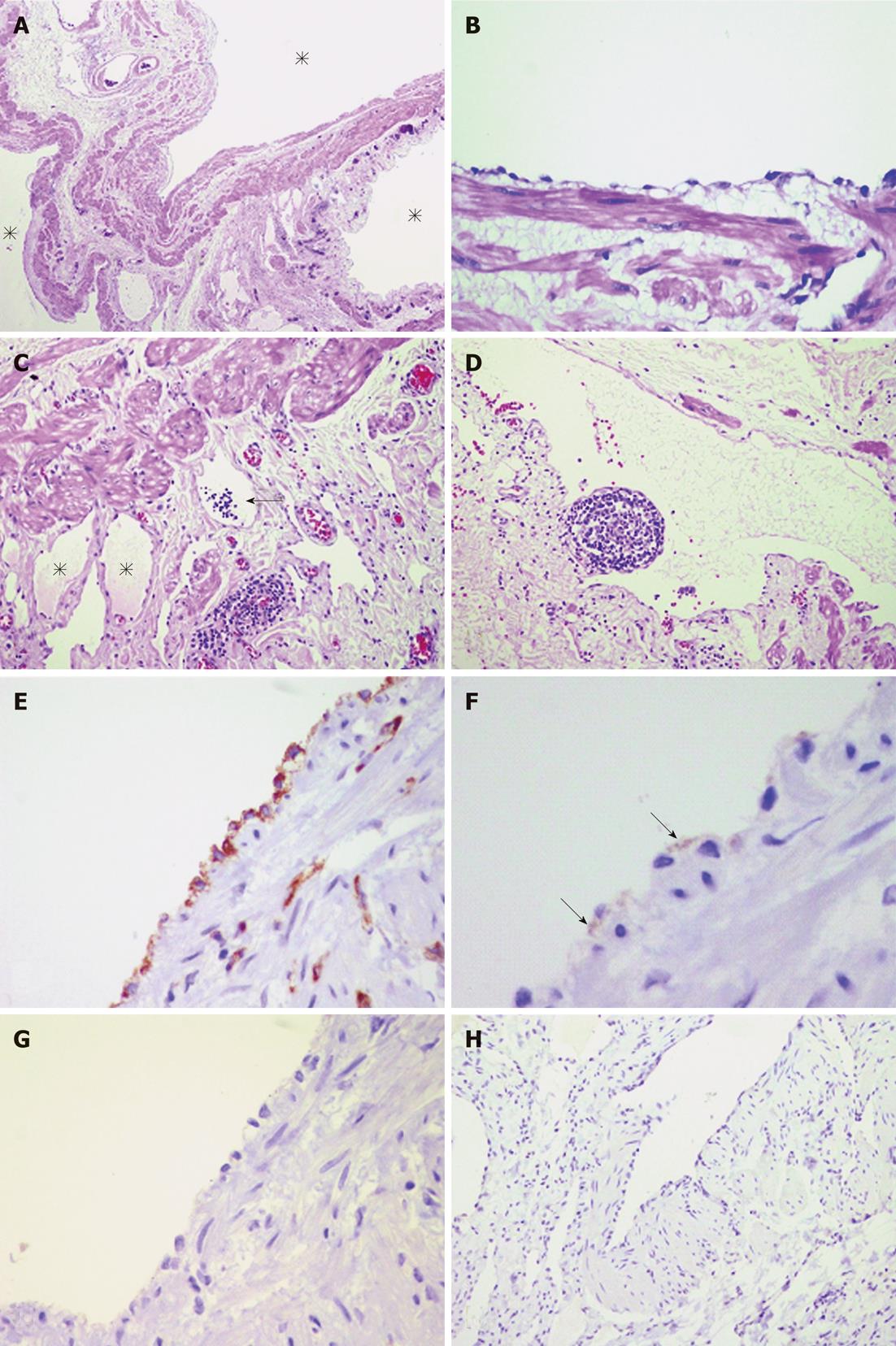
Figure 3 Histopathology.
A: Cystic spaces (asterisks) with smooth muscle in their walls (hematoxyin and eosin, 40 ×); B: Flat lining cells of the cystic spaces with smooth muscle below them (hematoxyin and eosin, 400 ×); C: Lymphatic spaces in the stroma containing lymphoid cells (arrow) and pale eosinophilic fluid, suggestive of lymph (asterisks) (hematoxyin and eosin, 200 ×); D: Subendothelial lymphoid follicle (hematoxyin and eosin, 200 ×); E-G: Immunohistochemical study of the lining cells of the cystic wall shows strong cytoplasmic immunoreactivity for factor VIII-related antigen (E, 600 ×) and focal reactivity with D2-40 (F, 1000 ×, arrows) but not for calretinin (G, 600 ×); H: Immunohistochemical study for human melanoma black-45 shows negative result in the smooth muscle of the lymphatic wall (100 ×).
- Citation: Suthiwartnarueput W, Kiatipunsodsai S, Kwankua A, Chaumrattanakul U. Lymphangioma of the small bowel mesentery: A case report and review of the literature. World J Gastroenterol 2012; 18(43): 6328-6332
- URL: https://www.wjgnet.com/1007-9327/full/v18/i43/6328.htm
- DOI: https://dx.doi.org/10.3748/wjg.v18.i43.6328


