Copyright
©2012 Baishideng Publishing Group Co.
World J Gastroenterol. Nov 21, 2012; 18(43): 6324-6327
Published online Nov 21, 2012. doi: 10.3748/wjg.v18.i43.6324
Published online Nov 21, 2012. doi: 10.3748/wjg.v18.i43.6324
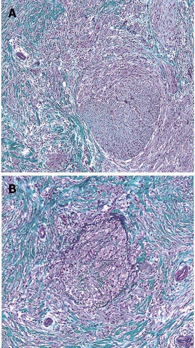
Figure 4 The granular cell tumor cells partially infiltrated a peripheral nerve fibrous tissue bunch in the bile duct wall.
In addition, they infiltrated the vein in the bile duct wall and occluded some of the vein lumina. A: Granular cell tumor involving a small nerve (Elastica-Masson stain, × 4); B: A vein is replaced by a neoplastic cell (Elastica-Masson stain, × 4).
- Citation: Saito J, Kitagawa M, Kusanagi H, Kano N, Ishii E, Nakaji S, Hirata N, Hoshi K. Granular cell tumor of the common bile duct: A Japanese case. World J Gastroenterol 2012; 18(43): 6324-6327
- URL: https://www.wjgnet.com/1007-9327/full/v18/i43/6324.htm
- DOI: https://dx.doi.org/10.3748/wjg.v18.i43.6324


