Copyright
©2012 Baishideng Publishing Group Co.
World J Gastroenterol. Nov 21, 2012; 18(43): 6216-6225
Published online Nov 21, 2012. doi: 10.3748/wjg.v18.i43.6216
Published online Nov 21, 2012. doi: 10.3748/wjg.v18.i43.6216
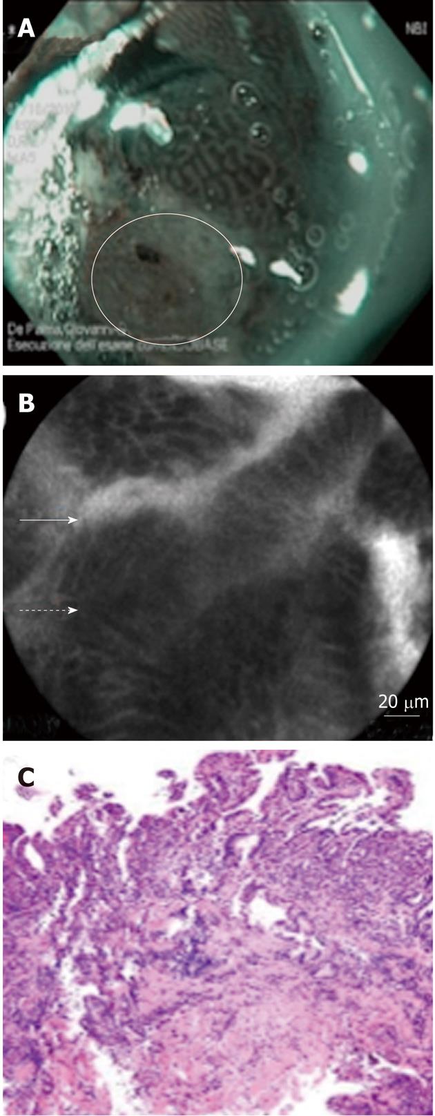
Figure 3 Enhanced narrow-band imaging and probe-based confocal laser endomicroscopy images of dysplastic Barrett’s esophagus.
A: Narrow-band imaging images shows distorted pits with irregular microvasculature (white circle); B: The corresponding probe-based confocal laser endomicroscopy image shows disorganized, distorted villiform structure and crypts, dark columnar cells (dash arrow) and dilated irregular vessels (solid arrow); C: High-grade dysplasia was found at histology in biopsy specimens performed at this level.
- Citation: Palma GDD. Management strategies of Barrett's esophagus. World J Gastroenterol 2012; 18(43): 6216-6225
- URL: https://www.wjgnet.com/1007-9327/full/v18/i43/6216.htm
- DOI: https://dx.doi.org/10.3748/wjg.v18.i43.6216


