Copyright
©2012 Baishideng Publishing Group Co.
World J Gastroenterol. Nov 14, 2012; 18(42): 6172-6176
Published online Nov 14, 2012. doi: 10.3748/wjg.v18.i42.6172
Published online Nov 14, 2012. doi: 10.3748/wjg.v18.i42.6172
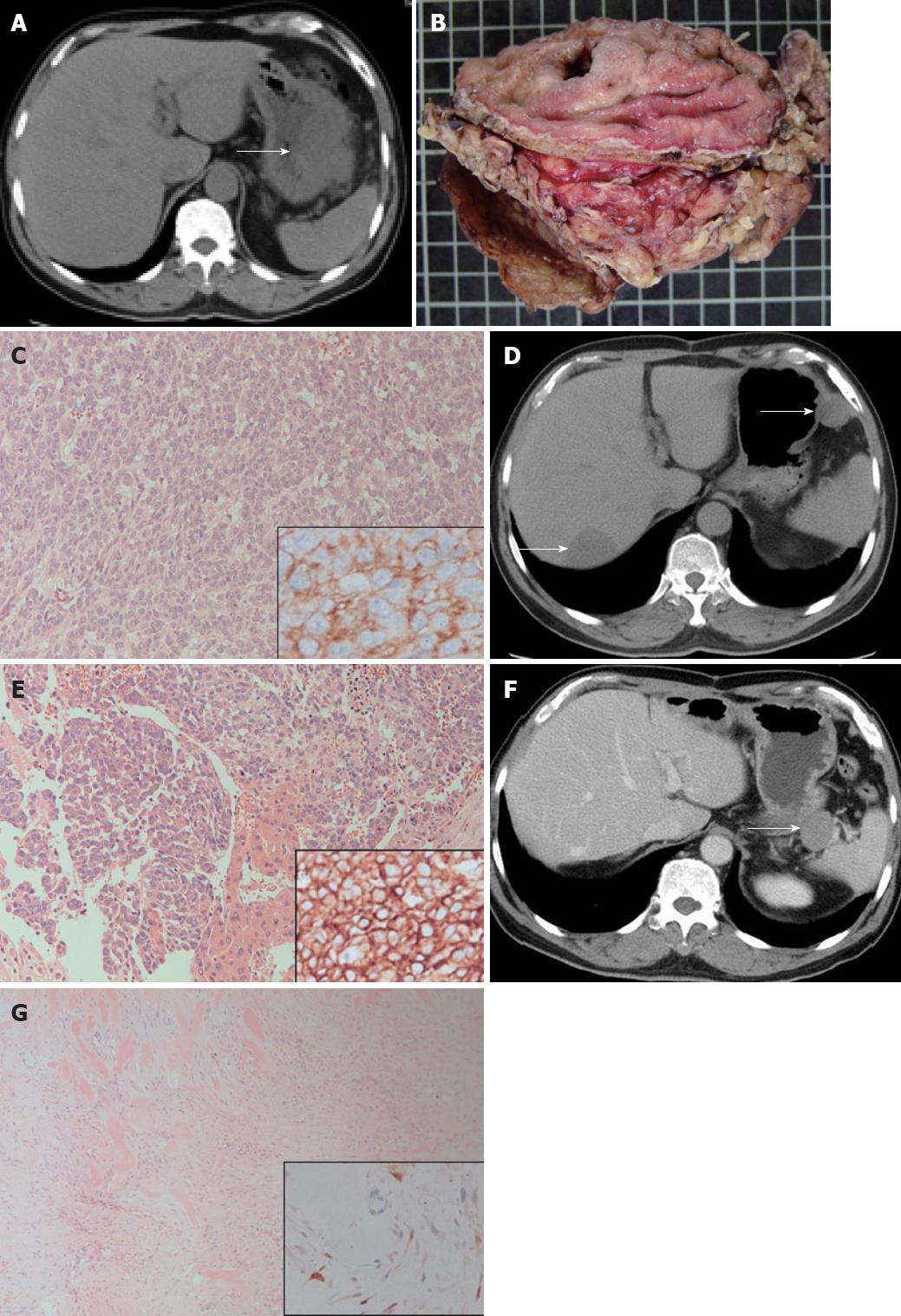
Figure 1 Abdominal computed tomography images and pathologic features of gastrointestinal stromal tumor, metastatic hepatic gastrointestinal stromal tumor and retroperitoneal desmoid tumor.
A: Abdominal computed tomography (CT) scan reveals a 10-cm tumor (arrow) located at the greater curvature of the stomach; B: The excised specimen discloses a patch of gastric mucosa with a central deep-seated ulcer and the gastrointestinal stromal tumor adhering to the red yellow omental tissues; C: Histologically, the 10-cm gastric tumor demonstrated sheets of CD117-positive epithelioid cells after immunohistochemical (IHC) staining (right lower inset), hematoxylin and eosin (HE) stain, ×200; D: The follow-up abdominal CT scan shows metastatic tumors in the liver (left lower arrow) and upper greater curvature of the stomach (right upper arrow); E: The histology of metastatic liver tumor demonstrated nests and sheets of CD117-positive epithelioid tumor cells (right lower inset, IHC stain). Entrapped hepatocytes are seen in the middle lower portion, HE stain, ×200; F: Abdominal CT scan discloses a tumor neogrowth (arrow) located in the retroperitoneum between the pancreatic tail and splenic hilar region; G: Histologically, the retroperitoneal tumor demonstrated proliferative spindle cells with keloid-like bundles and erythrocyte extravasation, HE stain, ×200. IHC staining reveals a positive nuclear beta-catenin in spindle cells (right lower inset).
- Citation: Shih LY, Wei CK, Lin CW, Tseng CE. Postoperative retroperitoneal desmoid tumor mimics recurrent gastrointestinal stromal tumor: A case report. World J Gastroenterol 2012; 18(42): 6172-6176
- URL: https://www.wjgnet.com/1007-9327/full/v18/i42/6172.htm
- DOI: https://dx.doi.org/10.3748/wjg.v18.i42.6172


